Stage 0 Perihilar Bile Duct Cancer Carcinoma In Situ
Stage 0 Perihilar Bile Duct Cancer Carcinoma In Situ represents the earliest, non-invasive form of bile duct cancer, confined to the innermost layer of the bile duct lining. Understanding this initial stage is crucial for timely intervention and improved patient outcomes.
Key Takeaways
- Stage 0 Perihilar Bile Duct Cancer Carcinoma In Situ is a non-invasive cancer, meaning abnormal cells are confined to the surface layer of the bile duct and have not spread.
- Due to its early nature, symptoms are often absent, making early diagnosis challenging and frequently incidental.
- Diagnosis typically involves advanced imaging techniques and endoscopic procedures to visualize the bile ducts and obtain tissue samples.
- Treatment primarily focuses on surgical removal of the affected bile duct segment, offering a high chance of cure.
- Regular surveillance is important after treatment to monitor for any recurrence or new developments.
What is Stage 0 Perihilar Bile Duct Cancer Carcinoma In Situ?
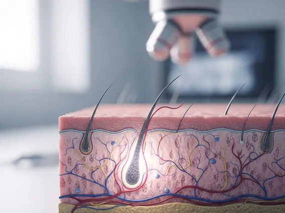
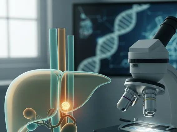
Stage 0 Perihilar Bile Duct Cancer Carcinoma In Situ refers to a very early form of cancer where abnormal cells are found only in the innermost layer of the bile duct lining and have not invaded deeper tissues or spread to other parts of the body. The term “perihilar” indicates that the cancer is located in the perihilar region, which is where the bile ducts from the liver merge before exiting the liver. This stage is considered non-invasive, meaning the cancerous cells are “in situ” (in place) and have not yet become invasive cancer. This early detection is rare, as bile duct cancers are often diagnosed at more advanced stages due to their subtle symptoms.
Bile duct cancers, also known as cholangiocarcinomas, are relatively uncommon. According to the American Cancer Society, approximately 8,000 people are diagnosed with cholangiocarcinoma each year in the United States, with perihilar types being the most common. While specific statistics for Stage 0 are not widely reported due to its rarity and often incidental discovery, identifying carcinoma in situ is critical for preventing progression to invasive disease.
Symptoms and Early Diagnosis of Perihilar Bile Duct Carcinoma In Situ
Due to its non-invasive nature, Carcinoma in situ perihilar bile duct symptoms are often absent or very subtle, making early detection particularly challenging. When symptoms do occur, they are typically non-specific and may include mild jaundice (yellowing of the skin or eyes), unexplained fatigue, itching, dark urine, or light-colored stools. However, these symptoms are more commonly associated with blockages caused by more advanced tumors, rather than Stage 0 disease.
The importance of Early stage perihilar bile duct cancer diagnosis cannot be overstated, as it significantly improves the prognosis. Diagnosis often occurs incidentally during investigations for other conditions or through advanced imaging techniques. Diagnostic procedures may include:
- Imaging Studies: Techniques such as magnetic resonance cholangiopancreatography (MRCP), computed tomography (CT) scans, and ultrasound can visualize the bile ducts and surrounding structures to identify any abnormalities.
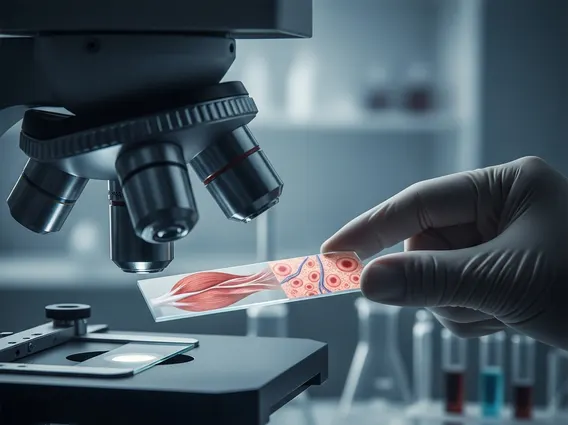
- Endoscopic Procedures: Endoscopic retrograde cholangiopancreatography (ERCP) or percutaneous transhepatic cholangiography (PTC) allow direct visualization of the bile ducts and can be used to collect tissue samples (biopsies) for pathological examination.
- Cholangioscopy: A procedure where a thin, flexible scope is inserted directly into the bile ducts to visually inspect the lining and obtain targeted biopsies, which is crucial for confirming carcinoma in situ.
These diagnostic tools help clinicians determine the exact location and extent of the abnormal cells, guiding subsequent treatment decisions.
Treatment Options for Perihilar Bile Duct Carcinoma In Situ
The primary goal of Perihilar bile duct carcinoma in situ treatment is the complete removal of the abnormal cells to prevent progression to invasive cancer. Given that the disease is confined to the superficial layer, treatment is often highly effective and potentially curative. The standard approach involves surgical resection.
Surgical options typically include:
- Bile Duct Resection: Removal of the affected segment of the bile duct.
- Partial Hepatectomy: If the carcinoma in situ extends into the liver tissue adjacent to the bile ducts, a portion of the liver might also be removed.
- Lymphadenectomy: Removal of nearby lymph nodes, although less common for pure carcinoma in situ, it might be performed to ensure no microscopic spread.
The extent of surgery depends on the precise location and spread of the carcinoma in situ. Following successful surgical removal, patients typically undergo regular surveillance, which may include follow-up imaging and blood tests, to monitor for any signs of recurrence or new lesions. The prognosis for Stage 0 perihilar bile duct cancer is generally excellent with complete surgical resection, highlighting the critical importance of early detection and intervention.