Somatostatin Receptor Scintigraphy
Somatostatin Receptor Scintigraphy is a diagnostic imaging technique used in nuclear medicine. It plays a crucial role in identifying and monitoring certain types of neuroendocrine tumors that express somatostatin receptors.
Key Takeaways
- Somatostatin Receptor Scintigraphy (SRS) is a nuclear medicine scan that detects tumors expressing somatostatin receptors.
- It involves injecting a radioactive tracer that binds to these receptors, allowing tumors to be visualized.
- The procedure is vital for diagnosing, staging, and monitoring neuroendocrine tumors (NETs).
- SRS helps guide treatment decisions, including peptide receptor radionuclide therapy (PRRT).
- Results indicate the presence, location, and extent of disease, aiding in prognosis and management.
What is Somatostatin Receptor Scintigraphy (SRS)?
Somatostatin Receptor Scintigraphy (SRS) is a specialized nuclear medicine imaging technique. It is primarily used to detect and localize neuroendocrine tumors (NETs) and other conditions that express somatostatin receptors on their cell surfaces. These receptors are specific proteins that bind to somatostatin, a naturally occurring hormone. By using a radioactive analog of somatostatin, medical professionals can visualize these tumors. The scan provides functional information about the tumor’s biological activity, which is crucial for diagnosis and treatment planning.
This diagnostic tool is particularly valuable because many NETs, such as carcinoid tumors, gastrinomas, and insulinomas, overexpress somatostatin receptors. The ability to image these receptors allows for precise localization of primary tumors and metastatic lesions, even those that might be difficult to detect with conventional imaging methods like CT or MRI. The information gained from SRS helps clinicians understand the extent of the disease and make informed decisions regarding patient management.
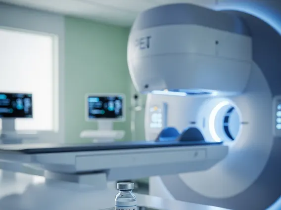
The Somatostatin Receptor Scintigraphy Procedure
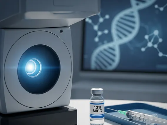
The somatostatin receptor scintigraphy procedure typically involves several steps, beginning with the administration of a radiopharmaceutical. Patients receive an intravenous injection of a tracer, most commonly an analog of somatostatin labeled with a radioactive isotope, such as Indium-111 (¹¹¹In-pentetreotide, often known as Octreoscan). This tracer travels through the bloodstream and selectively binds to somatostatin receptors present on tumor cells.
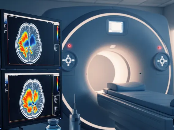
After the injection, there is a waiting period, usually ranging from 4 to 24 hours, to allow the tracer to accumulate in the target tissues and for background activity to clear from non-target areas. Imaging is then performed using a gamma camera, which detects the radiation emitted by the tracer. Multiple scans may be taken over one or two days to capture optimal images. The imaging process itself is non-invasive and generally well-tolerated.
- Preparation: Patients may be advised to fast for a few hours before the scan and to discontinue certain medications that could interfere with tracer uptake.
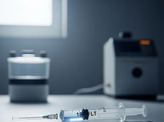
- Tracer Injection: A small amount of the radioactive somatostatin analog is injected into a vein.
- Waiting Period: Time is allowed for the tracer to circulate and bind to receptors, typically 4-24 hours.
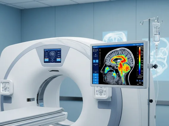
- Imaging: A specialized gamma camera captures images of the body, identifying areas where the tracer has accumulated. This may involve whole-body scans and/or SPECT (Single-Photon Emission Computed Tomography) for more detailed 3D images.
- Post-Scan: Patients are encouraged to drink plenty of fluids to help excrete the tracer from their system.
Interpreting Somatostatin Receptor Scintigraphy Results and Uses
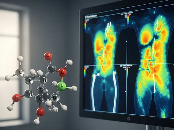
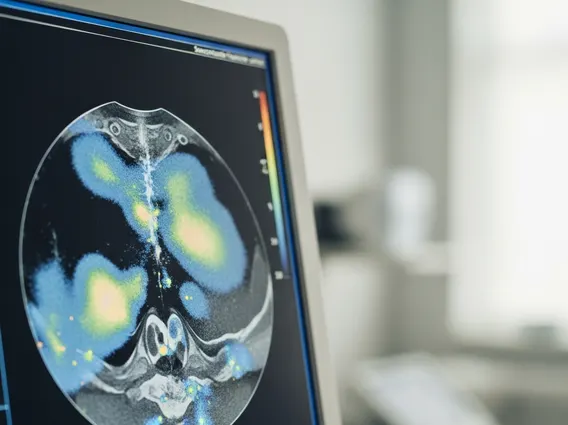
Somatostatin receptor scintigraphy results explained provide critical insights into the presence, location, and extent of somatostatin receptor-positive tumors. A positive result indicates areas where the radiotracer has accumulated, suggesting the presence of tumor cells expressing these receptors. The intensity of tracer uptake can also offer an indication of the receptor density on the tumor cells. Radiologists and nuclear medicine physicians interpret these images, often correlating them with other diagnostic tests to provide a comprehensive diagnosis.
The somatostatin receptor scintigraphy uses are extensive in the management of neuroendocrine tumors. It is invaluable for:
- Diagnosis: Confirming the presence of NETs when suspected based on symptoms or biochemical markers.
- Staging: Determining the primary tumor’s location and identifying any metastatic spread throughout the body.
- Treatment Planning: Guiding therapeutic decisions, particularly for patients who may benefit from somatostatin analog therapy or peptide receptor radionuclide therapy (PRRT), which targets these same receptors.
- Monitoring: Assessing treatment response and detecting disease recurrence or progression over time.
- Prognosis: Providing information that can help predict the tumor’s behavior and patient outcomes.
For instance, a study published in the Journal of Nuclear Medicine highlighted the utility of SRS in identifying primary tumors and metastases in patients with well-differentiated neuroendocrine tumors, often leading to changes in management plans. The ability of SRS to visualize tumors that might be occult on other imaging modalities makes it an indispensable tool in oncology.