Sestamibi Breast Imaging
Sestamibi Breast Imaging is a specialized nuclear medicine procedure used to evaluate breast tissue, particularly in cases where standard mammography may be inconclusive or challenging. This imaging technique offers valuable insights into cellular activity, aiding in the detection and characterization of breast lesions.
Key Takeaways
- Sestamibi Breast Imaging utilizes a radioactive tracer to highlight metabolically active breast tissue.
- It is particularly beneficial for women with dense breast tissue, which can obscure abnormalities on mammograms.
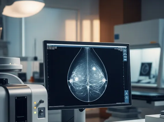
- The procedure involves an intravenous injection of the tracer followed by imaging with a specialized gamma camera.
- This scan aids in differentiating between benign and malignant breast lesions based on distinct tracer uptake patterns.
- It serves as a complementary diagnostic tool alongside other imaging methods like mammography and ultrasound in breast cancer assessment.
What is Sestamibi Breast Imaging?
Sestamibi Breast Imaging, also known as Molecular Breast Imaging (MBI) or Breast Scintigraphy, is a diagnostic technique that employs a small amount of a radioactive tracer called Technetium-99m Sestamibi. After intravenous injection, this tracer circulates throughout the body. Cancer cells, which typically exhibit higher metabolic activity and blood flow compared to normal cells, tend to absorb and retain more of the Sestamibi tracer. A specialized gamma camera then detects the gamma rays emitted by the tracer, producing detailed images that highlight areas of increased metabolic activity within the breast. This method is especially valuable for identifying suspicious areas that may be difficult to visualize with conventional mammography, particularly in women with dense breast tissue, where tumors can be masked by overlapping normal tissue. The primary goal is to provide a more precise assessment of breast abnormalities and to support the early detection of breast cancer.
The Sestamibi Breast Imaging Procedure: How It Works
The Sestamibi breast imaging procedure is typically an outpatient process that involves several distinct steps. Patients generally do not require extensive preparation, such as fasting, before the scan. The procedure begins with the intravenous injection of a small dose of the Technetium-99m Sestamibi tracer, usually into a vein in the arm. Following the injection, there is a brief waiting period, typically ranging from a few minutes to an hour, to allow the tracer to circulate and be adequately absorbed by breast tissue. Subsequently, the patient lies down on a specialized imaging table, and the breast is carefully positioned between two detectors of a gamma camera. The camera then captures images from various angles, detecting the gamma rays emitted by the tracer. These images reveal areas where the tracer has accumulated, indicating regions of higher metabolic activity. The entire imaging process usually takes approximately 45 to 60 minutes. A nuclear medicine physician or radiologist then meticulously interprets these images to identify any abnormal patterns of tracer uptake, which can suggest the presence of cancerous tissue.
Sestamibi Scans for Breast Cancer Detection
Sestamibi scans for breast cancer play a significant role in diagnostic pathways, particularly for specific patient populations. One of its most notable advantages is its enhanced effectiveness in women with dense breast tissue. Dense breasts contain a higher proportion of fibrous and glandular tissue compared to fatty tissue, which can appear white on a mammogram, potentially obscuring tumors that also present as white areas. Sestamibi imaging, by detecting metabolic activity rather than tissue density, can effectively overcome this limitation. According to the American Cancer Society, approximately 40% of women in the U.S. have dense breasts, underscoring the importance of complementary imaging tools. This scan aids in differentiating between benign and malignant lesions by revealing characteristic patterns of tracer uptake; malignant tumors typically demonstrate higher and more rapid uptake and retention of the tracer. While not a substitute for biopsy, a Sestamibi scan can guide further diagnostic steps and provide crucial information when other imaging results are inconclusive, thereby enhancing the accuracy of breast cancer detection and management.