Sertoli Leydig Cell Tumor
Sertoli Leydig Cell Tumor (SLCT) is a rare type of ovarian tumor that originates from the sex cord-stromal cells of the ovary. These tumors are notable for their ability to produce androgens, leading to masculinizing effects in affected individuals.

Key Takeaways
- Sertoli Leydig Cell Tumor is a rare ovarian tumor originating from sex cord-stromal cells.
- It often produces male hormones (androgens), leading to symptoms of virilization.
- Diagnosis involves clinical evaluation, hormone level measurements, and imaging studies.
- Surgical removal of the tumor is the primary and most effective treatment.
- Prognosis is generally favorable, especially for tumors diagnosed at an early stage.
What is Sertoli Leydig Cell Tumor?
Sertoli Leydig Cell Tumor is a rare neoplasm of the ovary, accounting for less than 0.5% of all ovarian cancers. These tumors are typically benign but can occasionally be malignant. They are characterized by their cellular composition, which resembles the Sertoli and Leydig cells found in the testes, and their endocrine activity. The tumor’s ability to produce androgens often leads to distinct clinical signs, making early detection possible in many cases. While they can occur at any age, they are most commonly diagnosed in young women and adolescents, with a median age of diagnosis around 25 years, according to various epidemiological studies.
Clinical Manifestations and Diagnosis of Sertoli Leydig Cell Tumor
The clinical presentation of SLCT is largely driven by its hormonal activity. The most prominent Sertoli Leydig cell tumor symptoms are those related to virilization, which results from the excessive production of androgens. These symptoms can develop gradually or rapidly and may include:
- Hirsutism: excessive growth of coarse body hair in a male pattern.
- Deepening of the voice: due to changes in the vocal cords.
- Clitoromegaly: enlargement of the clitoris.
- Menstrual irregularities: ranging from oligomenorrhea (infrequent periods) to amenorrhea (absence of periods).
- Acne and temporal balding: also linked to increased androgen levels.
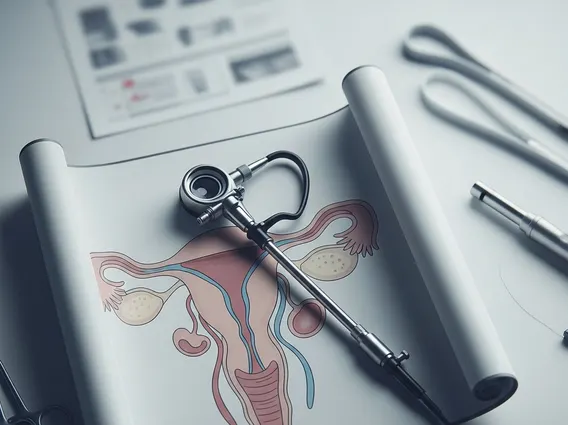
The Sertoli Leydig cell tumor diagnosis typically begins with a thorough physical examination and a review of the patient’s medical history, focusing on the onset and progression of virilizing symptoms. Blood tests are crucial for measuring hormone levels, particularly testosterone, which is often significantly elevated. Imaging studies, such as pelvic ultrasound, CT scans, or MRI, are then used to locate the ovarian mass and assess its size and characteristics. While imaging can strongly suggest the presence of an ovarian tumor, a definitive diagnosis requires a pathological examination of tissue obtained through surgery or biopsy.
Treatment Approaches for Sertoli Leydig Cell Tumor
The primary and most effective approach for Sertoli Leydig cell tumor treatment options is surgical removal of the tumor. The extent of surgery depends on several factors, including the patient’s age, desire for future fertility, the tumor’s stage, and whether it is unilateral or bilateral. For young women who wish to preserve fertility and have a unilateral, early-stage tumor, a unilateral oophorectomy (removal of the affected ovary) is often performed. In cases where fertility preservation is not a concern, or for more advanced tumors, a total hysterectomy with bilateral salpingo-oophorectomy (removal of the uterus, fallopian tubes, and ovaries) may be recommended.
The role of adjuvant therapies, such as chemotherapy or radiation therapy, is generally limited for SLCTs due to their rarity and typically good prognosis after surgical resection. These treatments are usually reserved for cases with malignant features, advanced stage, recurrence, or tumors that have spread beyond the ovary. Regular follow-up with imaging and hormone level monitoring is essential after treatment to detect any potential recurrence early. The overall prognosis for patients with Sertoli Leydig Cell Tumors is generally favorable, especially for those with well-differentiated tumors confined to the ovary.