Scintimammography
Scintimammography is a specialized nuclear medicine imaging technique employed in the detection and evaluation of breast cancer. It offers a functional assessment of breast tissue, complementing anatomical imaging methods like mammography by highlighting areas of increased metabolic activity often associated with malignant tumors.
Key Takeaways
- Scintimammography is a nuclear medicine test used to detect breast cancer, particularly in complex cases.
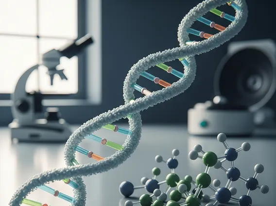
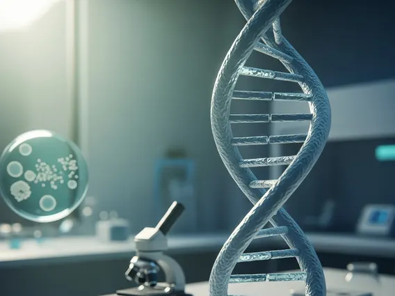
- It utilizes a radioactive tracer that accumulates in metabolically active cancer cells, providing functional information.
- The procedure involves an intravenous injection of a tracer, followed by imaging with a specialized gamma camera.
- It serves as a valuable supplementary tool to mammography, especially for women with dense breast tissue or challenging lesions.
- Unlike mammography, which provides structural images, scintimammography focuses on the biological activity of breast lesions.
What is Scintimammography?
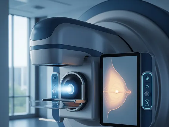
Scintimammography is a diagnostic imaging test that uses a small amount of a radioactive tracer to identify cancerous cells in the breast. This nuclear medicine technique is often employed when conventional mammography results are inconclusive, or for patients with dense breast tissue, which can make tumor detection difficult on standard X-ray mammograms. It provides functional information about breast lesions by detecting areas of increased blood flow and metabolic activity, which are characteristic features of malignant tumors.
This imaging modality is particularly useful for assessing the extent of known breast cancer, evaluating multifocal or multicentric disease, and monitoring response to neoadjuvant chemotherapy. It is not typically used as a primary screening tool but rather as a complementary diagnostic test to enhance the accuracy of breast cancer detection and staging.
Scintimammography Procedure: How It Works
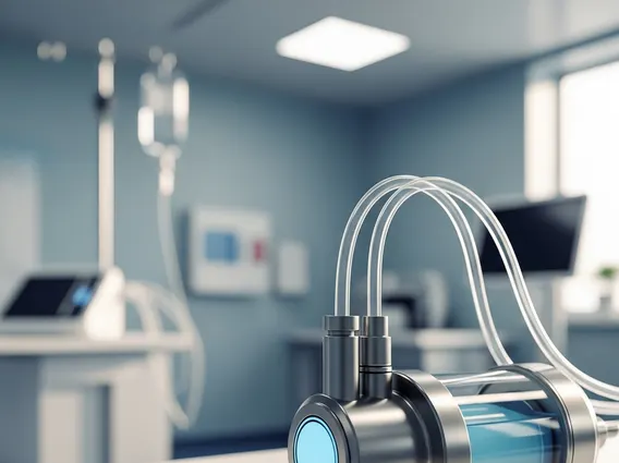
The scintimammography procedure details involve several key steps designed to visualize metabolically active breast lesions. The process begins with the intravenous injection of a radioactive tracer, most commonly technetium-99m sestamibi. This tracer is absorbed by cells throughout the body, but it tends to accumulate in areas with higher metabolic rates, such as rapidly growing cancer cells, more than in normal tissue.
After a short waiting period, typically 5 to 10 minutes to allow the tracer to distribute, the patient is positioned on a special imaging table. A gamma camera then captures images of the breasts. The patient may be asked to lie in various positions, such as prone with the breast hanging freely, to optimize image acquisition and ensure comprehensive coverage. The camera detects the gamma rays emitted by the tracer, creating detailed images that highlight areas where the tracer has accumulated. This is precisely how scintimammography works: by identifying these “hot spots” of tracer uptake, radiologists can pinpoint suspicious lesions that may indicate the presence of breast cancer. The entire imaging process usually takes about 30 to 60 minutes.
Scintimammography vs. Mammography: Key Differences
Understanding the scintimammography vs mammography differences is crucial for appreciating their respective roles in breast cancer diagnosis. While both are vital tools, they operate on distinct principles and provide different types of information. Mammography is an X-ray imaging technique that provides anatomical images of breast tissue, primarily looking for structural abnormalities like masses, calcifications, or architectural distortions. It is the gold standard for breast cancer screening.
Scintimammography, on the other hand, is a nuclear medicine technique that offers functional information. It assesses the metabolic activity of breast lesions, making it particularly effective in identifying rapidly growing tumors that may not be clearly visible on mammograms, especially in dense breasts. According to the American Cancer Society, approximately 40-50% of women have dense breast tissue, which can obscure tumors on mammograms, making supplementary imaging like scintimammography valuable in such cases.
The table below summarizes the key distinctions between these two important breast imaging modalities:
| Feature | Mammography | Scintimammography |
|---|---|---|
| Imaging Principle | X-ray imaging (anatomical structure) | Nuclear medicine (metabolic function) |
| Primary Use | Primary screening, diagnostic follow-up | Supplementary diagnostic tool, especially for dense breasts or inconclusive mammograms |
| What it Detects | Structural changes (masses, calcifications, distortions) | Areas of increased metabolic activity (tracer uptake) |
| Tracer/Contrast | No tracer; sometimes uses contrast for specific types (e.g., contrast-enhanced mammography) | Radioactive tracer (e.g., technetium-99m sestamibi) |
| Radiation Exposure | Ionizing radiation (X-rays) | Ionizing radiation (gamma rays from tracer) |
| Compression Required | Yes, breast compression is standard | No breast compression required |