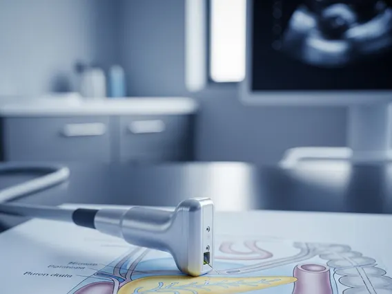
Endoscopic Ultrasound
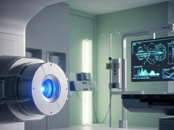
Endoscopic Ultrasound (EUS) is a sophisticated medical imaging technique that combines endoscopy with ultrasound technology to provide highly detailed images of internal organs and structures. This minimally invasive procedure plays a crucial role in diagnosing and staging various conditions within the gastrointestinal tract and surrounding areas.
Key Takeaways
- Endoscopic Ultrasound (EUS) combines endoscopy and ultrasound for detailed internal imaging.
- It offers superior visualization of the gastrointestinal tract walls and adjacent organs compared to traditional imaging.
- The procedure involves inserting a thin, flexible scope with an ultrasound transducer through the mouth or rectum.
- EUS is highly effective for diagnosing cancers, pancreatitis, and bile duct stones, and for guiding biopsies.
- It provides higher resolution and more precise staging capabilities than CT or MRI for certain conditions.
What is Endoscopic Ultrasound (EUS)?
Endoscopic Ultrasound (EUS) is a diagnostic procedure that merges two advanced medical technologies: endoscopy and ultrasound. It involves the use of a thin, flexible tube called an endoscope, which has a small ultrasound transducer at its tip. This allows physicians to obtain high-resolution ultrasound images from inside the body, specifically from within the gastrointestinal (GI) tract. The proximity of the ultrasound probe to the target organs, such as the pancreas, bile ducts, and GI tract walls, enables the capture of exceptionally clear and detailed images that are often superior to those obtained by external ultrasound or other imaging modalities for specific conditions.
The primary purpose of EUS is to visualize the layers of the GI tract wall, as well as adjacent organs like the pancreas, liver, gallbladder, and lymph nodes. This technique is invaluable for detecting abnormalities, assessing the extent of diseases, and guiding minimally invasive procedures. When considering what is Endoscopic Ultrasound, it is important to understand its ability to provide real-time, cross-sectional images, which aids in precise diagnosis and staging of various medical conditions.
Endoscopic Ultrasound Procedure and Benefits
The endoscopic ultrasound procedure explained typically begins with the patient receiving sedation to ensure comfort throughout the examination. The endoscope, equipped with an ultrasound transducer, is then gently guided through the mouth (for upper GI tract examinations) or rectum (for lower GI tract examinations) to the area of interest. As the endoscope moves, the ultrasound transducer emits sound waves that bounce off internal structures, creating detailed images displayed on a monitor. The physician can then meticulously examine the tissue layers and surrounding organs for any abnormalities.
The endoscopic ultrasound uses and benefits are extensive, making it a powerful tool in modern medicine. Some key applications and advantages include:
- Cancer Staging: EUS is highly effective in determining the depth of tumor invasion in the esophagus, stomach, rectum, and pancreas, and assessing involvement of nearby lymph nodes. This precise staging is critical for treatment planning.
- Diagnosis of Pancreatic and Bile Duct Conditions: It can detect small tumors, cysts, and stones in the pancreas and bile ducts that might be missed by other imaging tests.
- Biopsy Guidance: EUS allows for fine-needle aspiration (FNA) or biopsy of suspicious lesions or lymph nodes, providing tissue samples for pathological examination with high accuracy and minimal invasiveness.
- Evaluation of Submucosal Lesions: It helps differentiate between benign and malignant lesions located beneath the lining of the GI tract.
- Therapeutic Interventions: EUS can guide therapeutic procedures, such as draining cysts or injecting medications.
The minimally invasive nature of EUS, combined with its diagnostic precision, significantly reduces the need for more invasive surgical procedures for diagnosis, leading to faster recovery times and reduced risks for patients.
EUS Compared to Other Imaging Techniques
When considering endoscopic ultrasound vs other imaging modalities, EUS offers distinct advantages, particularly for conditions affecting the gastrointestinal tract and adjacent organs. While techniques like Computed Tomography (CT) scans, Magnetic Resonance Imaging (MRI), and traditional external ultrasound are valuable, EUS provides a unique perspective and level of detail due to the proximity of the ultrasound probe to the target area.
For instance, traditional external ultrasound can be limited by overlying bowel gas or obesity, which can obscure views of deep abdominal organs. CT and MRI scans provide excellent anatomical overviews but may not offer the same high-resolution detail of the GI tract wall layers or small lesions in the pancreas and bile ducts that EUS can achieve. EUS excels in visualizing the depth of tumor invasion, which is crucial for accurate cancer staging, and in detecting very small lesions that might be beyond the resolution of CT or MRI. Furthermore, EUS allows for real-time, image-guided biopsies, which is a capability not typically offered by CT or MRI without additional, separate procedures.
Here is a comparison of EUS with some other common imaging techniques:
| Feature | Endoscopic Ultrasound (EUS) | CT Scan | MRI Scan | Traditional External Ultrasound |
|---|---|---|---|---|
| Resolution of GI Wall/Adjacent Organs | Excellent (high detail, depth of invasion) | Good (overall anatomy) | Good (soft tissue contrast) | Variable (limited by gas/depth) |
| Ability to Guide Biopsy | Yes (real-time, precise FNA) | Yes (but often less precise for small lesions) | No (typically not real-time for biopsy) | Yes (for superficial lesions) |
| Detection of Small Lesions | Superior (especially in pancreas/bile duct) | Good | Good | Limited (especially deep lesions) |
| Invasiveness | Minimally invasive (sedation required) | Non-invasive (radiation exposure) | Non-invasive (no radiation) | Non-invasive (no radiation) |
| Primary Use Case | Detailed GI wall/pancreatic/biliary assessment, cancer staging, biopsy | Broad anatomical survey, emergency imaging | Soft tissue characterization, neurological imaging | Initial screening, superficial organ assessment |
While each imaging technique has its strengths, EUS stands out for its unique ability to provide intricate details of the GI tract and surrounding structures, making it an indispensable tool for specific diagnostic challenges and therapeutic interventions.