Ekg
An Ekg, commonly known as an electrocardiogram, is a vital and non-invasive medical test used to assess the electrical activity of the heart. It provides essential insights into cardiac rhythm, rate, and overall heart health, playing a crucial role in the diagnosis and management of various cardiovascular conditions.
Key Takeaways
- An EKG records the heart’s electrical signals, offering a detailed view of its rhythm and function.
- The procedure involves placing electrodes on the skin to detect the electrical impulses generated by the heart muscle.
- EKGs are instrumental in diagnosing a wide range of heart conditions, including arrhythmias, signs of heart attacks, and structural issues.
- Interpretation focuses on analyzing specific waveforms (P, QRS, T) and intervals to identify patterns indicative of cardiac abnormalities.
- This quick, painless, and widely accessible diagnostic tool is fundamental in modern cardiology for evaluating symptoms and monitoring heart health.
What is an EKG (Electrocardiogram)?
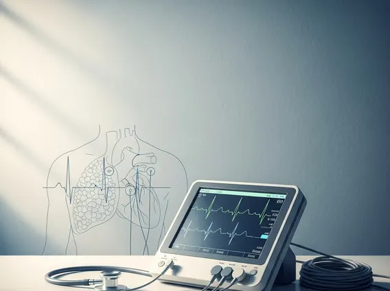
An Ekg, or electrocardiogram, is a diagnostic test that measures and records the electrical activity of the heart over a period of time. This electrical activity is what triggers the heart muscle to contract and relax, enabling it to pump blood efficiently throughout the body. By capturing these electrical impulses, an EKG produces a detailed graphical representation of the heart’s rhythm, rate, and the strength and timing of its electrical signals. It serves as a cornerstone diagnostic tool in cardiology, offering a non-invasive method to evaluate cardiac function. The information derived from an EKG is invaluable for healthcare professionals in detecting and characterizing a broad spectrum of heart conditions, making it a routine procedure in many clinical settings.
How an EKG Test Works
The procedure for an EKG test is straightforward, quick, and entirely painless. During the test, several small, adhesive electrodes are carefully placed on specific areas of the patient’s chest, arms, and legs. These electrodes are connected by wires to an EKG machine, which acts as a sensitive receiver for the heart’s electrical signals. As the heart beats, it generates minute electrical currents that propagate through the body. The electrodes detect these currents, and the EKG machine amplifies and translates them into a series of characteristic waves that are either displayed on a monitor in real-time or printed onto a grid paper. The entire recording process typically lasts only a few minutes, providing a comprehensive snapshot of the heart’s electrical performance at that precise moment. This allows clinicians to observe the heart’s electrical axis, rhythm, rate, and the integrity of its conduction pathways.
Purpose and Basic Interpretation of an EKG
The primary purpose of EKG test is to diagnose and monitor a variety of heart conditions, making it an indispensable tool in cardiovascular medicine. It is frequently used to identify abnormalities in heart rhythm, known as arrhythmias, detect evidence of a heart attack (myocardial infarction) or past heart damage, assess the efficacy of certain cardiac medications, and screen for structural issues such as an enlarged heart or inflammation of the heart muscle. EKG interpretation basics involve a systematic analysis of the distinct waveforms (P wave, QRS complex, T wave) and the intervals between them, which are displayed on the EKG tracing.
- The P wave signifies the electrical depolarization and contraction of the atria, the heart’s upper chambers.
- The QRS complex represents the rapid electrical depolarization and contraction of the ventricles, the powerful lower chambers responsible for pumping blood to the body and lungs.
- The T wave indicates the electrical repolarization and relaxation of the ventricles, preparing them for the next beat.
By meticulously examining the morphology, duration, and amplitude of these waves, as well as the lengths of the intervals and segments, healthcare providers can pinpoint deviations from normal cardiac electrical activity. For example, an unusually fast, slow, or irregular rhythm can indicate an arrhythmia, while specific changes in the ST segment (the segment between the S wave and T wave) are often tell-tale signs of myocardial ischemia or infarction. An EKG serves as a critical initial diagnostic step when patients present with symptoms like chest pain, shortness of breath, dizziness, or palpitations, guiding subsequent diagnostic tests and therapeutic interventions.