Hematoxylin And Eosin Staining
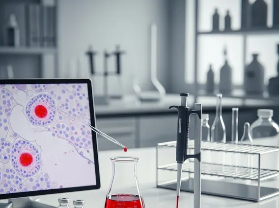
Hematoxylin And Eosin (H&E) staining is a foundational technique in histology, indispensable for visualizing tissue morphology and diagnosing various medical conditions. This method provides critical insights into cellular structures and tissue architecture.

Key Takeaways
- Hematoxylin And Eosin (H&E) staining is a widely used histological technique for examining tissue samples.
- It employs two dyes: hematoxylin, which stains cell nuclei blue/purple, and eosin, which stains cytoplasm and extracellular matrix pink/red.
- The primary purpose of H&E stain is to reveal the detailed microscopic anatomy of tissues, aiding in the diagnosis of diseases.
- The H&E staining principle relies on the differential binding of acidic and basic dyes to cellular components.
- The multi-step hematoxylin and eosin staining procedure involves tissue processing, sectioning, and sequential application of the dyes.
What is Hematoxylin And Eosin (H&E) Staining?
Hematoxylin And Eosin (H&E) staining refers to a fundamental and widely utilized histological technique in medical diagnostics and research. It is a cornerstone method for pathologists to examine tissue samples under a microscope, providing a clear visual representation of cellular components and tissue organization. This technique is crucial for identifying normal tissue structures, detecting pathological changes, and diagnosing a broad spectrum of diseases, including cancers, inflammatory conditions, and degenerative disorders.
The purpose of H&E stain is to differentiate various tissue components based on their chemical properties. By imparting distinct colors to different parts of the cell and extracellular matrix, H&E staining allows for the detailed assessment of tissue architecture, cellular morphology, and the presence of any abnormal cells or structures. This clarity is essential for accurate diagnosis and for guiding treatment decisions in clinical settings.
Principle and Procedure of H&E Staining
The H&E staining principle is based on the interaction of two dyes with different cellular and extracellular components. Hematoxylin is a basic dye that stains basophilic (acid-loving) structures, primarily the cell nuclei, which contain nucleic acids (DNA and RNA), a deep blue or purple. Eosin, an acidic dye, stains acidophilic (base-loving) structures, such as the cytoplasm, collagen, and muscle fibers, various shades of pink or red. This differential staining creates a high-contrast image, allowing pathologists to easily distinguish between different cell types and tissue elements.
The hematoxylin and eosin staining procedure involves several critical steps to prepare tissue samples for microscopic examination. These steps ensure the tissue is properly preserved, sectioned, and stained to reveal its intricate details. A typical procedure includes:
- Fixation: Preserving tissue structure and preventing degradation, often using formalin.
- Processing: Dehydrating the tissue through alcohol gradients, clearing it with xylene, and embedding it in paraffin wax for support.
- Sectioning: Cutting the paraffin-embedded tissue into very thin slices (typically 3-5 micrometers) using a microtome.
- Deparaffinization and Rehydration: Removing the paraffin wax and rehydrating the tissue sections for aqueous staining.
- Hematoxylin Staining: Immersing sections in hematoxylin solution to stain nuclei blue/purple.
- Rinsing and Bluing: Washing off excess hematoxylin and treating with a bluing agent (e.g., dilute ammonia water) to enhance the blue color.
- Eosin Staining: Immersing sections in eosin solution to stain cytoplasm and extracellular matrix pink/red.
- Dehydration, Clearing, and Mounting: Dehydrating the stained sections, clearing them in xylene, and mounting them under a coverslip with a permanent mounting medium for long-term preservation and viewing.
Each step in the procedure is meticulously performed to ensure optimal staining quality, which is paramount for accurate diagnostic interpretation by pathologists.