Mediastinum
The mediastinum is a crucial anatomical compartment within the chest, housing vital organs and structures. Understanding its components and potential pathologies is fundamental in medical diagnostics and treatment.
Key Takeaways
- The mediastinum is the central compartment of the thoracic cavity, located between the lungs.
- It contains essential organs such as the heart, great vessels, trachea, esophagus, and thymus.
- Anatomically, it is divided into superior and inferior (anterior, middle, posterior) sections.
- Its primary function is to protect and support these vital structures while allowing their necessary movements.
- Various conditions, including tumors, infections, and cysts, can affect the mediastinum, often requiring precise diagnosis.
What is the Mediastinum?
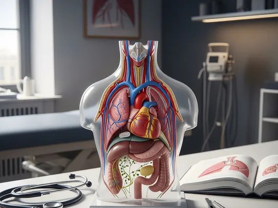
The what is the mediastinum refers to the central compartment of the thoracic cavity, situated between the two pleural sacs that enclose the lungs. It extends vertically from the thoracic inlet (superiorly) to the diaphragm (inferiorly), and horizontally from the sternum (anteriorly) to the vertebral column (posteriorly). This vital space is densely packed with numerous essential organs and structures, making it a critical area for both normal physiological function and potential disease processes. Its strategic position means that conditions affecting the mediastinum can have widespread implications for respiratory, cardiovascular, and digestive systems.
Anatomy, Divisions, and Function
The mediastinum anatomy and function are complex, as it houses a multitude of critical structures. For descriptive purposes, the mediastinum is traditionally divided into superior and inferior parts by an imaginary transverse plane passing from the sternal angle anteriorly to the intervertebral disc between the fourth and fifth thoracic vertebrae posteriorly. The inferior mediastinum is further subdivided into anterior, middle, and posterior compartments.
- Superior Mediastinum: Contains the thymus, trachea, esophagus, great vessels (aortic arch, brachiocephalic veins, superior vena cava), and nerves (vagus, phrenic, recurrent laryngeal).
- Anterior Mediastinum: The smallest division, located between the sternum and the pericardium. It primarily contains loose connective tissue, lymph nodes, and the lower part of the thymus in children.
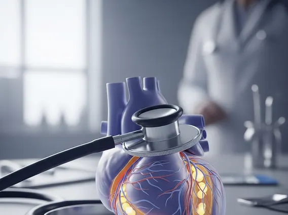
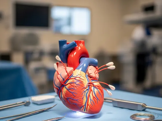
- Middle Mediastinum: The most important subdivision, containing the heart, pericardium, roots of the great vessels, main bronchi, and phrenic nerves.
- Posterior Mediastinum: Located behind the pericardium and diaphragm, containing the esophagus, descending thoracic aorta, azygos and hemiazygos veins, thoracic duct, and sympathetic trunks.
The mediastinum location in body is central to its function, which is primarily to protect and provide a pathway for vital structures connecting the neck, thorax, and abdomen. It allows for the passage of air, food, and blood, while also housing the primary organ responsible for circulating blood throughout the body. The loose connective tissue within the mediastinum also allows for movement of organs like the heart and esophagus during their respective functions.
Common Mediastinal Conditions
A variety of common mediastinal conditions can arise due to the diverse range of tissues and organs within this compartment. These conditions can be broadly categorized into masses (tumors, cysts), infections, and inflammatory processes. Mediastinal tumors are among the most frequently encountered pathologies, with their prevalence varying by age and specific location within the mediastinum. For instance, thymomas are common in the anterior mediastinum, while neurogenic tumors often occur in the posterior mediastinum.
According to a review published in the Journal of Thoracic Disease, primary mediastinal tumors are relatively rare, with an estimated incidence of 1-10 cases per 100,000 population per year, but they represent a significant diagnostic challenge due to their varied presentations.
Other conditions include:
- Mediastinitis: Inflammation of the mediastinum, often a severe infection resulting from esophageal perforation or post-surgical complications.
- Mediastinal Cysts: Non-cancerous fluid-filled sacs, such as bronchogenic cysts, pericardial cysts, or enteric cysts, which are usually congenital.
- Lymphadenopathy: Enlarged lymph nodes, which can be indicative of infection, inflammation, or malignancy (e.g., lymphoma, metastatic cancer).
Diagnosis typically involves imaging techniques like chest X-rays, CT scans, and MRI, often followed by biopsy for definitive characterization. Early detection and accurate diagnosis are crucial for effective management of these conditions.