Bone Scintigraphy
Bone scintigraphy is a diagnostic imaging technique used to detect and diagnose a variety of bone conditions. It provides detailed information about bone metabolism and blood flow, revealing problems not always visible on standard X-rays.
Key Takeaways
- Bone scintigraphy uses a small amount of radioactive tracer to create images of your bones.
- It helps identify bone abnormalities like fractures, infections, and tumors by showing areas of altered bone activity.
- The procedure involves an injection of the tracer, followed by imaging several hours later.
- It is a safe and effective tool for diagnosing a wide range of skeletal issues.
What is Bone Scintigraphy?
What is bone scintigraphy? Also known as a bone scan, this is a nuclear medicine imaging test that helps diagnose and track several types of bone diseases. It involves injecting a small amount of radioactive material, called a tracer, into a vein. This tracer travels through the bloodstream and accumulates in areas of the bone undergoing rapid metabolic activity or repair. The images produced reveal these areas, providing crucial insights into bone health and detecting issues like fractures, infections, and cancer that might not be visible on conventional X-rays in their early stages.
How Bone Scintigraphy Works
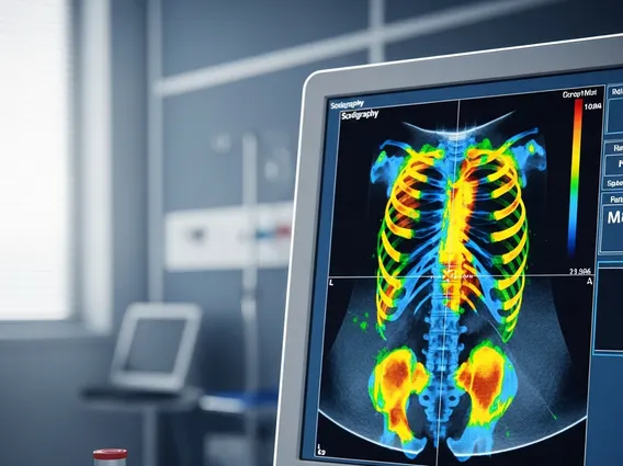
How does bone scintigraphy work? This diagnostic technique relies on areas of bone with increased metabolic activity absorbing more of the injected radioactive tracer. Once injected, the tracer, typically technetium-99m-labeled bisphosphonate, travels through the bloodstream and binds to areas of new bone formation. After a few hours, allowing the tracer to accumulate and unbound tracer to be cleared, a special camera called a gamma camera scans the body. This camera detects the gamma rays emitted by the tracer, creating detailed images that highlight areas of abnormal bone activity. These “hot spots” indicate potential issues, while “cold spots” might suggest areas with reduced blood flow or certain types of tumors.
Preparing for Your Bone Scan
Patients typically receive specific instructions from their healthcare provider. Generally, minimal preparations are required, but it’s important to:
- Inform your doctor about any medications, allergies, or if you are pregnant or breastfeeding.
- Drink plenty of water between the injection and the scan to help clear any unbound tracer from your system.
- Avoid calcium supplements or certain medications before the scan, if advised by your doctor.
What Happens During the Scan
The bone scan procedure explanation involves two main stages. First, a small amount of radioactive tracer is injected into a vein, usually in the arm. After the injection, there’s typically a waiting period of 2-4 hours, allowing the tracer to circulate and accumulate in the bones while excess tracer is naturally cleared. During the second stage, you will lie on a table while a gamma camera moves slowly over your body, capturing images. The scan itself is painless and usually takes 30-60 minutes. Sometimes, a “three-phase bone scan” is performed, involving initial images immediately after injection, followed by images a few minutes later, and then the delayed images, to assess blood flow and soft tissue uptake.
Post-Scan Information
After the scan, you can typically resume your normal activities immediately. The small amount of radioactive tracer used will naturally decay and be eliminated from your body over a few days, primarily through urine. Drinking extra fluids can help expedite this process. Your doctor will review the images and discuss the findings with you, often comparing them with previous imaging studies if available.
Uses of Bone Scintigraphy
The uses of bone scintigraphy are extensive, making it a valuable tool in various medical specialties. It is highly sensitive in detecting changes in bone metabolism, often identifying problems earlier than conventional X-rays. Key applications include:
- Detecting fractures: Especially stress fractures or occult fractures not visible on standard X-rays.
- Diagnosing bone infections (osteomyelitis): Identifying inflammation and infection in the bone, crucial for timely treatment.
- Identifying bone tumors and metastases: Bone scans are highly effective in detecting the spread of cancer to the bones (metastases) from primary tumors elsewhere. According to the American Cancer Society, bone metastases are common, occurring in up to 70% of patients with advanced breast and prostate cancer.
- Assessing arthritis and other inflammatory bone conditions: Helping to determine the extent and activity of conditions like rheumatoid arthritis.
- Evaluating unexplained bone pain: When the cause of bone pain is unclear, a bone scan can help pinpoint the source.
- Monitoring bone graft viability: Assessing the success of bone grafts after surgery.
This tool is critical for guiding treatment and monitoring disease progression for a wide array of skeletal conditions.