Bone Mineral Density Scan
A Bone Mineral Density (BMD) scan is a crucial diagnostic tool used to assess bone health. It provides valuable insights into bone strength and helps identify conditions like osteoporosis before fractures occur.
Key Takeaways
- A BMD scan, often a DEXA scan, measures bone mineral density to assess bone strength.
- The primary purpose of bone mineral density test is to diagnose osteoporosis and evaluate fracture risk.
- How bone density scans are performed typically involves a quick, non-invasive procedure where the patient lies on a table while a scanner passes over specific body areas.
- Interpreting bone density scan results relies on T-scores and Z-scores, which compare a patient’s bone density to healthy young adults or age-matched peers.
- Early detection through a bone mineral density scan allows for timely interventions to manage bone loss and reduce fracture risk.
What is a Bone Mineral Density Scan?
A Bone Mineral Density Scan, commonly known as a DEXA (Dual-energy X-ray Absorptiometry) scan, is a non-invasive medical test that measures the amount of calcium and other minerals in your bones. This measurement helps determine bone strength and density. The primary purpose of bone mineral density test is to diagnose osteoporosis, a condition characterized by weak and brittle bones, and to assess an individual’s risk of future fractures. It is a key diagnostic tool for monitoring bone health over time, especially in individuals at higher risk. According to the International Osteoporosis Foundation, worldwide, osteoporosis causes more than 8.9 million fractures annually, resulting in an osteoporotic fracture every 3 seconds. Early detection through a BMD scan is vital for timely intervention.
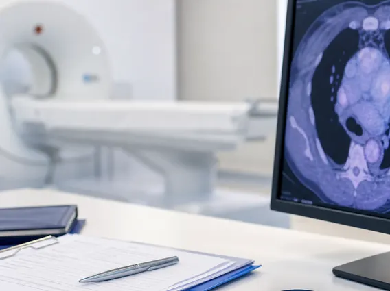
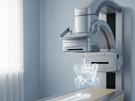
How Bone Density Scans Are Performed
Understanding how bone density scans are performed can help alleviate any concerns about the procedure. The most common type of BMD scan is the DEXA scan, which uses a very low dose of X-rays to measure bone density, typically in the hip and spine, as these areas are most indicative of overall bone health and are common sites for osteoporotic fractures. During the scan, the patient lies comfortably on a padded table. A mechanical arm then passes over the body, sending two different X-ray beams through the bones. The scanner calculates the bone density based on how much of each X-ray beam is absorbed by the bone and soft tissue. The entire procedure is quick, usually taking only 10-20 minutes, and is painless. Patients are advised to wear loose, comfortable clothing and avoid wearing items with metal fasteners, zippers, or buttons, as these can interfere with the X-ray images.
Interpreting Bone Density Scan Results
After the scan, a radiologist analyzes the images and provides a report, which a healthcare provider then uses for interpreting bone density scan results. The results are typically reported using two main scores: T-scores and Z-scores.
- T-score: This score compares your bone density to that of a healthy young adult of the same sex.
- A T-score of -1.0 or above is considered normal bone density.
- A T-score between -1.0 and -2.5 indicates osteopenia, meaning lower than normal bone density but not yet osteoporosis.
- A T-score of -2.5 or below indicates osteoporosis.
- Z-score: This score compares your bone density to that of other people in your age group, sex, and ethnic background. A very low Z-score might suggest that there are other factors contributing to bone loss beyond normal aging, such as an underlying medical condition or medication.
It is crucial to discuss these results with your doctor, as they will consider your overall health history, risk factors, and other clinical information to provide a comprehensive diagnosis and recommend appropriate management strategies, which may include lifestyle changes, medication, or further tests.