Avascular Necrosis
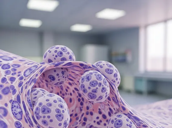
Avascular necrosis (AVN), also known as osteonecrosis, is a serious condition characterized by the death of bone tissue due to a lack of blood supply. This interruption can lead to tiny breaks in the bone and, eventually, to the bone’s collapse.
Key Takeaways
- AVN results from impaired blood flow to bone tissue, leading to its death and potential collapse.
- Symptoms often include joint pain that worsens over time, particularly in the hip, knee, or shoulder.
- Common causes range from trauma and steroid use to alcohol abuse and certain medical conditions.
- Diagnosis typically involves imaging tests like X-rays, MRI, and CT scans.
- Treatment aims to preserve the joint and can include medication, physical therapy, or surgery.
What is Avascular Necrosis?
Avascular necrosis (AVN), also known as osteonecrosis, is a serious condition where bone tissue dies due to an inadequate blood supply. This interruption deprives bone cells of oxygen and nutrients, leading to their demise and weakening the bone structure. Over time, this cellular death can cause tiny fractures and eventual collapse of the affected bone, damaging the adjacent joint. While AVN can affect any bone, it most commonly impacts the ends of long bones like the femur head in the hip, the knee, and the shoulder. Understanding what is avascular necrosis is crucial for recognizing its potential impact on joint health and mobility.
Avascular Necrosis: Symptoms and Causes
Understanding the signs and underlying factors of avascular necrosis is crucial for early detection and intervention. While AVN progression varies, distinct patterns of avascular necrosis symptoms causes have been identified.
Recognizing AVN Symptoms
In its early stages, avascular necrosis often presents with no symptoms. As the condition progresses and more bone tissue is affected, pain becomes the predominant symptom. The location of pain depends on the affected bone; for instance, hip AVN often causes pain in the groin, thigh, or buttock, while knee AVN results in pain in the knee area.
- Joint Pain: Initially, pain may only occur when putting weight on the affected joint. Over time, it can become constant, even at rest.
- Limited Range of Motion: The affected joint may become stiff, and its ability to move freely can decrease.
- Limping: If the hip or knee is affected, a noticeable limp may develop due to pain and joint dysfunction.
- Gradual Onset: Symptoms typically develop gradually over weeks or months.
Common Causes of Avascular Necrosis
A variety of factors can disrupt blood flow to bones, leading to avascular necrosis. While some cases are idiopathic (of unknown cause), several risk factors are well-established. Corticosteroid use and alcohol abuse are significant risk factors, accounting for a notable percentage of non-traumatic AVN cases.
- Trauma: Fractures or dislocations near a joint can damage blood vessels, interrupting blood supply to the bone.
- Corticosteroid Use: Long-term, high-dose use of corticosteroids (e.g., prednisone) is a significant risk factor.
- Alcohol Abuse: Excessive alcohol consumption can lead to fatty deposits in blood vessels, reducing blood flow.
- Sickle Cell Disease: This genetic blood disorder can cause blockages in small blood vessels.
- Gaucher’s Disease: A rare genetic disorder causing fatty substances to build up in organs and bones.
- Radiation Therapy and Chemotherapy: These cancer treatments can sometimes damage blood vessels and bone tissue.
- Decompression Sickness: Affecting divers, this condition can lead to gas bubbles blocking blood flow.
- Organ Transplantation: Patients often receive high doses of corticosteroids, increasing their risk.
Diagnosing and Treating Avascular Necrosis
Early and accurate diagnosis is critical for effective management of avascular necrosis, as timely intervention can prevent or delay joint replacement surgery. Treatment strategies are tailored to the individual, considering disease stage, affected bone, and overall health. Both diagnosing avascular necrosis and understanding avascular necrosis treatment options are key to managing this condition effectively.
How AVN is Diagnosed
Diagnosing avascular necrosis typically involves a combination of physical examination, patient history, and advanced imaging studies. MRI’s ability to detect AVN early is crucial, allowing intervention before significant bone collapse.
- Physical Examination: A doctor will assess joint tenderness, range of motion, and any pain during movement.
- X-rays: In early stages, X-rays may appear normal. As the disease progresses, they can show bone changes, such as flattening of the bone or joint space narrowing.
- Magnetic Resonance Imaging (MRI): MRI is the most sensitive imaging test for diagnosing AVN, capable of detecting the condition in its very early stages, often before symptoms appear or X-rays show changes.
- Computed Tomography (CT) Scan: CT scans can provide more detailed information about the extent of bone damage and collapse.
- Bone Scan: A nuclear medicine test that can identify areas of increased or decreased bone metabolism, indicating AVN.
Treatment Options for Avascular Necrosis
The goal of avascular necrosis treatment is to improve joint function, reduce pain, and prevent further bone damage. Treatment choice is highly individualized, aiming to preserve the natural joint as long as possible.
Non-Surgical Treatments (Early Stages):
- Medications: Pain relievers, NSAIDs, cholesterol-lowering drugs (to reduce fatty deposits), and blood thinners (to improve blood flow) may be prescribed.
- Physical Therapy: Exercises to maintain or improve joint range of motion and strength.
- Rest: Reducing weight-bearing activities on the affected joint can help slow progression.
- Electrical Stimulation: Some studies suggest it may encourage new bone growth.
Surgical Treatments (Advanced Stages or Failed Non-Surgical):
- Core Decompression: A surgeon removes a section of bone from the inner layer, reducing pressure and creating channels for new blood vessels. This is most effective in early stages before bone collapse.
- Bone Grafting: Healthy bone tissue (from another part of the body or a donor) is transplanted to the affected area to support the joint and promote healing.
- Osteotomy: A wedge of bone is removed above or below a weight-bearing joint to shift weight to a healthier part of the bone.
- Joint Replacement: For severe cases where the joint has collapsed and conservative treatments have failed, total joint replacement (e.g., total hip replacement) may be necessary.