Diagnosis, Screening, and Early Detection of Neuroblastoma
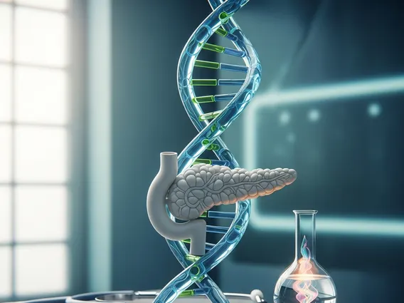
Neuroblastoma is a rare cancer that primarily affects infants and young children, developing from immature nerve cells found in several areas of the body, most commonly in the adrenal glands. Understanding the signs, diagnostic processes, and the critical role of early detection is paramount for improving outcomes for affected children.

Key Takeaways
- Neuroblastoma diagnosis often begins with subtle, non-specific symptoms that can vary depending on the tumor’s location.
- A comprehensive diagnostic journey involves initial medical evaluation, referral to specialists, and a combination of imaging, lab tests, and definitive biopsy.
- Key Neuroblastoma diagnosis methods include MIBG scans, CT/MRI, urine catecholamine tests, and pathological confirmation from a biopsy.
- While mass Neuroblastoma screening tests are not universally adopted, routine pediatric check-ups play a vital role in the Early detection of neuroblastoma.
- The Importance of early neuroblastoma diagnosis cannot be overstated, as it significantly impacts treatment options and overall prognosis.
Recognizing Early Signs of Neuroblastoma in Children
Neuroblastoma can present with a wide range of symptoms, often subtle and non-specific, making early recognition challenging. These Signs of neuroblastoma in children frequently mimic those of more common childhood illnesses, necessitating careful observation by parents and healthcare providers.
Common Symptoms by Location
The specific symptoms depend largely on where the tumor originates and whether it has spread. Common presentations include:
- Abdomen: The most common site, often presenting as a painless lump or swelling in the abdomen. Children may experience abdominal pain, constipation, or a feeling of fullness.
- Chest: Tumors in the chest can cause breathing difficulties, coughing, or Horner’s syndrome (drooping eyelid, small pupil, and decreased sweating on one side of the face).
- Neck: A visible lump in the neck, sometimes accompanied by nerve compression symptoms.
- Spinal Cord: If the tumor presses on the spinal cord, it can lead to weakness in the legs, difficulty walking, or changes in bowel and bladder function.
- Bone Marrow: Spread to the bone marrow can cause bone pain, limping, fever, fatigue, and paleness due to anemia.
- Eyes: Tumors around the eyes can cause dark circles (often called “raccoon eyes”), proptosis (bulging eyes), or vision changes.
- Skin: In infants, small, blue-red skin nodules may appear, which can blanch (turn white) when pressed.
- Systemic Symptoms: General symptoms like unexplained fever, weight loss, irritability, and persistent fatigue can also occur, indicating systemic involvement or tumor growth.
When to Seek Medical Attention
Parents should seek prompt medical attention if their child exhibits any persistent or worsening symptoms, especially if they are unusual for the child or do not resolve with typical care. While many symptoms can be attributed to benign conditions, a thorough evaluation by a pediatrician is crucial to rule out serious underlying issues, including neuroblastoma. Early consultation can be a critical step in the Early detection of neuroblastoma.
The Neuroblastoma Diagnosis Journey
When a child presents with suspicious symptoms, the path to a definitive neuroblastoma diagnosis involves several stages, often requiring collaboration among various medical specialists.
Initial Medical Evaluation
The diagnostic journey typically begins with a visit to the pediatrician. The doctor will take a detailed medical history, asking about the child’s symptoms, their duration, and any family history of cancer. A comprehensive physical examination will be performed to check for lumps, swelling, signs of pain, and overall health status. Based on these initial findings, the pediatrician may order preliminary tests, such as blood work or an ultrasound, to investigate the cause of the symptoms.
Referral to Specialists
If initial evaluations suggest the possibility of a serious condition like neuroblastoma, the child will be referred to a pediatric oncologist, a doctor specializing in childhood cancers. Other specialists, such as pediatric surgeons, radiologists, and pathologists, will also become integral to the diagnostic team. This multidisciplinary approach ensures a thorough investigation and accurate determination of How is neuroblastoma diagnosed, guiding subsequent treatment decisions.
Key Diagnostic Methods for Neuroblastoma
A definitive neuroblastoma diagnosis relies on a combination of advanced imaging techniques, specialized laboratory tests, and, most critically, tissue biopsy. These Neuroblastoma diagnosis methods help identify the tumor, assess its extent, and confirm its cellular characteristics.
Imaging Scans and Lab Tests
Various imaging modalities are used to locate the primary tumor and detect any spread:
- Ultrasound: Often the first imaging test for abdominal masses, providing a quick, non-invasive view.
- X-ray: Can reveal tumors in the chest or bone abnormalities.
- Computed Tomography (CT) Scan: Provides detailed cross-sectional images, useful for assessing tumor size, location, and involvement of surrounding structures.
- Magnetic Resonance Imaging (MRI): Offers superior soft tissue contrast, particularly valuable for evaluating tumors near the spinal cord or brain.
- Metaiodobenzylguanidine (MIBG) Scan: A specialized nuclear medicine scan where a radioactive tracer (MIBG) is injected. Neuroblastoma cells often absorb MIBG, making this scan highly effective for detecting primary tumors and metastatic sites throughout the body.
- Bone Scan: Used to check for cancer spread to the bones.
Laboratory tests are also crucial:
- Urine Catecholamine Tests: Neuroblastoma cells often produce high levels of catecholamines (hormones like adrenaline and noradrenaline) and their breakdown products, such as vanillylmandelic acid (VMA) and homovanillic acid (HVA). Measuring these substances in a 24-hour urine sample is a key diagnostic indicator.
- Blood Tests: Complete blood count (CBC) to check for anemia or other blood abnormalities, and tests for electrolytes and kidney/liver function.
Biopsy and Pathological Confirmation
While imaging and lab tests can strongly suggest neuroblastoma, a biopsy is essential for a definitive neuroblastoma diagnosis. A small sample of the tumor tissue is surgically removed and examined under a microscope by a pathologist. This examination confirms the presence of cancer cells, identifies the specific type of neuroblastoma, and helps determine its aggressiveness. Bone marrow biopsies are also frequently performed to check for cancer cells in the bone marrow, which indicates metastatic disease.
The table below summarizes common diagnostic tests and their primary purpose:
| Diagnostic Method | Primary Purpose |
|---|---|
| Physical Exam & History | Initial assessment of symptoms and general health. |
| Urine Catecholamine Tests (VMA/HVA) | Detect elevated tumor markers in urine. |
| CT/MRI Scans | Detailed imaging of tumor size, location, and spread. |
| MIBG Scan | Specific detection of neuroblastoma cells in primary and metastatic sites. |
| Biopsy (Tumor/Bone Marrow) | Definitive pathological confirmation and staging. |
Screening and Early Detection Approaches
The concept of mass Neuroblastoma screening tests has been explored, but their widespread implementation remains a subject of debate. Nevertheless, proactive approaches contribute significantly to the Early detection of neuroblastoma.
Urine Catecholamine Screening
In some countries, such as Japan, mass screening programs for neuroblastoma were implemented in the past, primarily using urine VMA/HVA tests for infants at 6 months of age. The rationale was that detecting tumors at an early, asymptomatic stage could lead to better outcomes. However, these programs have largely been discontinued in many regions, including the United States, due to concerns about overdiagnosis and overtreatment of tumors that might have regressed spontaneously without intervention, known as “silent” neuroblastomas. The American Academy of Pediatrics does not recommend routine screening for neuroblastoma in asymptomatic infants.
Role of Routine Check-ups
Despite the limitations of mass screening, routine pediatric check-ups remain a cornerstone for the Early detection of neuroblastoma. During these visits, pediatricians conduct physical examinations, monitor growth and development, and discuss any parental concerns. A skilled pediatrician may detect an abdominal mass or other subtle Signs of neuroblastoma in children during a routine exam, prompting further investigation. These regular visits provide opportunities for early intervention, even in the absence of specific screening tests.
The Critical Importance of Early Neuroblastoma Diagnosis
The Importance of early neuroblastoma diagnosis cannot be overstated, as it is a primary determinant of treatment success and long-term survival rates. Neuroblastoma is a highly heterogeneous disease, and its prognosis is strongly linked to the child’s age at diagnosis and the stage of the disease.
Children diagnosed at a younger age (typically under 18 months) and those with localized disease often have a significantly better prognosis compared to older children or those with metastatic disease. According to the American Cancer Society, the 5-year survival rate for neuroblastoma varies widely by stage and age, with localized disease having survival rates often above 90%, while high-risk, metastatic disease can have rates significantly lower. Early detection allows for treatment when the tumor is smaller, potentially less aggressive, and more amenable to surgical removal or less intensive chemotherapy regimens. This can lead to fewer side effects and a better quality of life for the child.
Conversely, a delayed neuroblastoma diagnosis can mean the cancer has progressed to a more advanced stage, potentially spreading to bones, bone marrow, or other organs. Advanced-stage neuroblastoma requires more aggressive and prolonged treatment, which may include high-dose chemotherapy, stem cell transplant, radiation therapy, and immunotherapy, all of which carry greater risks and side effects. Therefore, recognizing early signs and pursuing timely diagnostic evaluations are crucial steps in improving outcomes for children with this challenging cancer.
Frequently Asked Questions
What are the most common initial signs of neuroblastoma?
The initial signs of neuroblastoma are often subtle and non-specific, making them challenging to identify. Common indicators include a painless lump in the abdomen or neck, unexplained fever, persistent fatigue, irritability, bone pain, or changes in bowel habits. In some cases, children may develop dark circles around their eyes or small, blue-red skin nodules. Any persistent or unusual symptom in a child warrants medical evaluation to ensure early detection.
Why is a biopsy essential for diagnosing neuroblastoma?
While imaging scans and urine tests can strongly suggest neuroblastoma, a biopsy is crucial for a definitive diagnosis. It involves surgically removing a small tissue sample from the suspected tumor, which is then examined under a microscope by a pathologist. This examination confirms the presence of neuroblastoma cells, determines the specific type, and helps assess its aggressiveness, providing vital information for accurate staging and treatment planning.
Are there routine screening tests for neuroblastoma?
Currently, there are no universally recommended routine mass screening tests for neuroblastoma in asymptomatic infants in countries like the United States. While some regions historically implemented urine catecholamine screening, concerns about overdiagnosis of tumors that might regress spontaneously led to their discontinuation. Instead, regular pediatric check-ups are emphasized, as they provide opportunities for pediatricians to identify subtle signs during physical examinations and developmental assessments, aiding in early detection.