
Urothelial Cancer: Symptoms, Stages, Treatment Methods

Urothelial Cancer: Symptoms, Stages, Treatment Methods

Urothelial Cancer: Symptoms, Stages, Treatment Methods

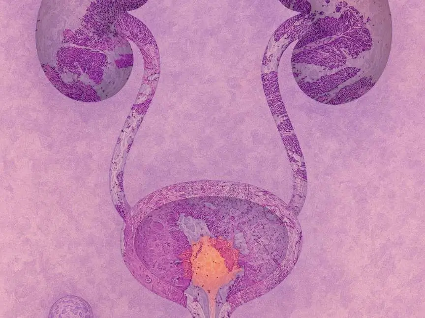
Urothelial cancer is a malignancy arising from the urothelium, the specialized epithelial tissue lining the bladder, ureters, renal pelvis, and urethra. It represents the most common form of bladder cancer and a significant global health concern, making early recognition and accurate staging essential for improving patient outcomes.

Key Takeaways

  • Urothelial cancer originates in the urothelial lining of the urinary tract and is most frequently found in the bladder.
  • Blood in the urine (hematuria) is the most common early warning sign and should never be ignored.
  • Staging ranges from non-muscle-invasive (Stage 0–I) to metastatic disease (Stage IV), guiding treatment decisions.
  • Treatment options include surgery, intravesical therapy, chemotherapy, immunotherapy, and targeted therapy depending on the stage.
  • Five-year survival rates vary dramatically by stage, reinforcing the importance of early detection and timely intervention.

Urothelial Cancer Symptoms and Early Warning Signs

Recognizing urothelial cancer symptoms and warning signs at the earliest possible stage can significantly improve the likelihood of successful treatment. The condition often presents with subtle urinary changes that patients may attribute to infection or aging, which can unfortunately delay diagnosis. Understanding which symptoms warrant immediate medical attention is therefore critical for anyone at elevated risk.

The most prevalent and clinically significant early symptom is hematuria, or blood in the urine. Blood may appear visibly, turning urine pink, red, or brownish, or it may be detected only through urinalysis (microscopic hematuria). According to the American Cancer Society, gross hematuria occurs in approximately 80–90% of bladder cancer cases, which represents the most common form of urothelial malignancy. Importantly, hematuria is often painless in its earliest occurrence, leading patients to dismiss it as temporary or benign.

Beyond hematuria, urothelial carcinoma early symptoms to know include persistent urinary urgency, increased frequency of urination, and a burning sensation during urination — symptoms that can closely mimic a urinary tract infection. Pelvic or lower back pain may develop as the tumor grows. In more advanced cases, patients may experience fatigue, unintentional weight loss, swelling in the lower limbs due to lymphatic involvement, and bone pain if metastasis has occurred. Any combination of these symptoms, particularly when they persist without a clear infectious cause, should prompt a prompt evaluation by a healthcare professional.

  • Blood in the urine (gross or microscopic hematuria)
  • Increased urinary frequency or urgency
  • Pain or burning sensation during urination
  • Lower abdominal or pelvic discomfort
  • Unexplained fatigue or weight loss (typically in advanced disease)
  • Flank or back pain if the upper urinary tract is involved

Certain risk factors increase the likelihood of developing this condition, including smoking — the single largest modifiable risk factor — as well as occupational exposure to aromatic amines found in the dye, rubber, and paint industries. Age above 55, male sex, and a personal or family history of urinary tract malignancies also elevate risk. Recognizing these factors alongside early symptoms helps guide timely screening decisions.

How Urothelial Cancer Is Diagnosed and Staged

Accurate diagnosis and staging are the foundation of effective disease management. Clinicians employ a combination of laboratory tests, imaging studies, and tissue evaluation to confirm the presence of malignancy and assess its extent. This multi-modal approach ensures that treatment planning is both precise and individualized.

The diagnostic process typically begins with a urinalysis and urine cytology, in which shed cells from the urinary tract are examined under a microscope for malignant characteristics. Cystoscopy — a procedure in which a thin, flexible camera is inserted into the bladder through the urethra — remains the gold standard for direct visualization of tumors. When a suspicious lesion is identified, a transurethral resection of bladder tumor (TURBT) is performed both to remove tissue for biopsy and, in some cases, as a therapeutic intervention. Imaging studies such as computed tomography (CT) urography, magnetic resonance imaging (MRI), and bone scans are used to assess the depth of tumor invasion and detect distant metastases.

Staging follows the TNM (Tumor, Node, Metastasis) classification system established by the American Joint Committee on Cancer (AJCC). This system evaluates how deeply the tumor has invaded surrounding tissue, whether regional lymph nodes are involved, and whether distant spread has occurred. The stage at diagnosis has a direct and substantial impact on prognosis and treatment selection, making thorough staging an indispensable step in clinical care.

Stage Description Invasion Level
Stage 0 (Ta/Tis) Non-invasive papillary carcinoma or carcinoma in situ Confined to the inner lining
Stage I (T1) Tumor invades connective tissue beneath the epithelium Lamina propria only
Stage II (T2) Muscle-invasive disease Muscularis propria
Stage III (T3–T4a) Extension beyond the bladder wall Perivesical fat or adjacent organs
Stage IV (T4b/N+/M1) Involvement of pelvic or abdominal wall, lymph nodes, or distant organs Metastatic spread

Molecular and genomic testing is increasingly used alongside traditional staging, particularly for identifying specific alterations such as FGFR3 mutations or PD-L1 expression levels. These biomarkers help oncologists select targeted or immunotherapy-based regimens, reflecting a broader shift toward precision oncology in the management of this disease.

Urothelial Cancer Treatment Options and Survival Rates

Urothelial cancer treatment options explained across the disease spectrum reveal a diverse therapeutic landscape shaped by tumor stage, grade, patient health status, and molecular profile. Treatment strategies range from bladder-preserving approaches for early-stage disease to aggressive systemic regimens for advanced or metastatic cases.

For non-muscle-invasive disease (Stages 0 and I), TURBT is the cornerstone of initial management. Following resection, intravesical therapy — in which medication is instilled directly into the bladder — is commonly used to reduce recurrence risk. Bacillus Calmette-Guérin (BCG) immunotherapy is the preferred intravesical agent for high-risk non-muscle-invasive tumors, as it stimulates a local immune response against residual cancer cells. Intravesical chemotherapy with agents such as mitomycin C may be used in lower-risk settings. Regular surveillance cystoscopy is essential following treatment, as recurrence rates for non-muscle-invasive disease can exceed 50% within five years.

Muscle-invasive urothelial cancer (Stage II–III) typically requires radical cystectomy — surgical removal of the bladder — combined with neoadjuvant cisplatin-based chemotherapy administered before surgery to improve long-term outcomes. In select patients, a bladder-sparing trimodality approach combining maximal TURBT, chemotherapy, and radiation therapy offers a viable alternative. For patients ineligible for cisplatin, carboplatin-based regimens or immunotherapy agents such as pembrolizumab may be considered.

Metastatic urothelial cancer (Stage IV) is managed primarily with systemic therapy. First-line treatment typically involves platinum-based combination chemotherapy, with gemcitabine plus cisplatin or gemcitabine plus carboplatin being standard regimens. Immune checkpoint inhibitors — including atezolizumab and pembrolizumab — have demonstrated durable responses in patients with platinum-ineligible disease or after disease progression. Additionally, erdafitinib, an FGFR inhibitor approved by the U.S. Food and Drug Administration (FDA), represents a targeted option for patients with specific FGFR2/3 alterations. Enfortumab vedotin, an antibody-drug conjugate, has also shown significant efficacy in previously treated metastatic disease.

Urothelial cancer treatment methods and survival rates are closely linked to disease stage at the time of diagnosis. According to the National Cancer Institute’s SEER database, the five-year relative survival rate for localized bladder cancer — which is predominantly urothelial in origin — exceeds 70%, while regional disease carries approximately a 38% five-year survival rate. For distant metastatic disease, the five-year survival rate falls to around 7–8%. These figures underscore why early detection and stage-appropriate intervention are so consequential.

Frequently Asked Questions

Can urothelial cancer be cured if detected early?

Yes, early-stage urothelial cancer confined to the inner lining of the bladder has a favorable prognosis and is often curable with TURBT and intravesical therapy. Stage 0 and Stage I tumors carry five-year survival rates well above 70% with appropriate treatment and surveillance. However, recurrence remains common even after successful initial therapy, making ongoing monitoring with regular cystoscopy an essential component of long-term care for all patients.

Is urothelial cancer the same as bladder cancer?

Urothelial cancer is the most common type of bladder cancer, accounting for approximately 90% of all bladder malignancies according to the American Cancer Society. However, not all urothelial cancers originate in the bladder — they can also arise in the renal pelvis, ureters, and urethra. The term “urothelial carcinoma” refers specifically to the cell type involved, while “bladder cancer” is a broader anatomical designation that can include other rarer histological subtypes.

What lifestyle changes can reduce the risk of developing urothelial cancer?

Smoking cessation is the most impactful lifestyle modification, as tobacco use accounts for approximately 50% of bladder cancer cases. Limiting exposure to occupational carcinogens such as aromatic amines, maintaining adequate hydration to reduce the concentration of urinary toxins, and attending routine medical check-ups when risk factors are present can further reduce risk. Individuals with a history of chronic urinary infections or prior pelvic radiation should also discuss personalized screening strategies with their healthcare provider.


[EN] Cancer Types
Cancer Clinical Trial Options

Specialized matching specifically for oncology clinical trials and cancer care research.

Your Birthday


By filling out this form, you're consenting only to release your medical records. You're not agreeing to participate in clinical trials yet.

Urothelial cancer is a malignancy arising from the urothelium, the specialized epithelial tissue lining the bladder, ureters, renal pelvis, and urethra. It represents the most common form of bladder cancer and a significant global health concern, making early recognition and accurate staging essential for improving patient outcomes.

Key Takeaways

  • Urothelial cancer originates in the urothelial lining of the urinary tract and is most frequently found in the bladder.
  • Blood in the urine (hematuria) is the most common early warning sign and should never be ignored.
  • Staging ranges from non-muscle-invasive (Stage 0–I) to metastatic disease (Stage IV), guiding treatment decisions.
  • Treatment options include surgery, intravesical therapy, chemotherapy, immunotherapy, and targeted therapy depending on the stage.
  • Five-year survival rates vary dramatically by stage, reinforcing the importance of early detection and timely intervention.

Urothelial Cancer Symptoms and Early Warning Signs

Recognizing urothelial cancer symptoms and warning signs at the earliest possible stage can significantly improve the likelihood of successful treatment. The condition often presents with subtle urinary changes that patients may attribute to infection or aging, which can unfortunately delay diagnosis. Understanding which symptoms warrant immediate medical attention is therefore critical for anyone at elevated risk.

The most prevalent and clinically significant early symptom is hematuria, or blood in the urine. Blood may appear visibly, turning urine pink, red, or brownish, or it may be detected only through urinalysis (microscopic hematuria). According to the American Cancer Society, gross hematuria occurs in approximately 80–90% of bladder cancer cases, which represents the most common form of urothelial malignancy. Importantly, hematuria is often painless in its earliest occurrence, leading patients to dismiss it as temporary or benign.

Beyond hematuria, urothelial carcinoma early symptoms to know include persistent urinary urgency, increased frequency of urination, and a burning sensation during urination — symptoms that can closely mimic a urinary tract infection. Pelvic or lower back pain may develop as the tumor grows. In more advanced cases, patients may experience fatigue, unintentional weight loss, swelling in the lower limbs due to lymphatic involvement, and bone pain if metastasis has occurred. Any combination of these symptoms, particularly when they persist without a clear infectious cause, should prompt a prompt evaluation by a healthcare professional.

  • Blood in the urine (gross or microscopic hematuria)
  • Increased urinary frequency or urgency
  • Pain or burning sensation during urination
  • Lower abdominal or pelvic discomfort
  • Unexplained fatigue or weight loss (typically in advanced disease)
  • Flank or back pain if the upper urinary tract is involved

Certain risk factors increase the likelihood of developing this condition, including smoking — the single largest modifiable risk factor — as well as occupational exposure to aromatic amines found in the dye, rubber, and paint industries. Age above 55, male sex, and a personal or family history of urinary tract malignancies also elevate risk. Recognizing these factors alongside early symptoms helps guide timely screening decisions.

How Urothelial Cancer Is Diagnosed and Staged

Accurate diagnosis and staging are the foundation of effective disease management. Clinicians employ a combination of laboratory tests, imaging studies, and tissue evaluation to confirm the presence of malignancy and assess its extent. This multi-modal approach ensures that treatment planning is both precise and individualized.

The diagnostic process typically begins with a urinalysis and urine cytology, in which shed cells from the urinary tract are examined under a microscope for malignant characteristics. Cystoscopy — a procedure in which a thin, flexible camera is inserted into the bladder through the urethra — remains the gold standard for direct visualization of tumors. When a suspicious lesion is identified, a transurethral resection of bladder tumor (TURBT) is performed both to remove tissue for biopsy and, in some cases, as a therapeutic intervention. Imaging studies such as computed tomography (CT) urography, magnetic resonance imaging (MRI), and bone scans are used to assess the depth of tumor invasion and detect distant metastases.

Staging follows the TNM (Tumor, Node, Metastasis) classification system established by the American Joint Committee on Cancer (AJCC). This system evaluates how deeply the tumor has invaded surrounding tissue, whether regional lymph nodes are involved, and whether distant spread has occurred. The stage at diagnosis has a direct and substantial impact on prognosis and treatment selection, making thorough staging an indispensable step in clinical care.

Stage Description Invasion Level
Stage 0 (Ta/Tis) Non-invasive papillary carcinoma or carcinoma in situ Confined to the inner lining
Stage I (T1) Tumor invades connective tissue beneath the epithelium Lamina propria only
Stage II (T2) Muscle-invasive disease Muscularis propria
Stage III (T3–T4a) Extension beyond the bladder wall Perivesical fat or adjacent organs
Stage IV (T4b/N+/M1) Involvement of pelvic or abdominal wall, lymph nodes, or distant organs Metastatic spread

Molecular and genomic testing is increasingly used alongside traditional staging, particularly for identifying specific alterations such as FGFR3 mutations or PD-L1 expression levels. These biomarkers help oncologists select targeted or immunotherapy-based regimens, reflecting a broader shift toward precision oncology in the management of this disease.

Urothelial Cancer Treatment Options and Survival Rates

Urothelial cancer treatment options explained across the disease spectrum reveal a diverse therapeutic landscape shaped by tumor stage, grade, patient health status, and molecular profile. Treatment strategies range from bladder-preserving approaches for early-stage disease to aggressive systemic regimens for advanced or metastatic cases.

For non-muscle-invasive disease (Stages 0 and I), TURBT is the cornerstone of initial management. Following resection, intravesical therapy — in which medication is instilled directly into the bladder — is commonly used to reduce recurrence risk. Bacillus Calmette-Guérin (BCG) immunotherapy is the preferred intravesical agent for high-risk non-muscle-invasive tumors, as it stimulates a local immune response against residual cancer cells. Intravesical chemotherapy with agents such as mitomycin C may be used in lower-risk settings. Regular surveillance cystoscopy is essential following treatment, as recurrence rates for non-muscle-invasive disease can exceed 50% within five years.

Muscle-invasive urothelial cancer (Stage II–III) typically requires radical cystectomy — surgical removal of the bladder — combined with neoadjuvant cisplatin-based chemotherapy administered before surgery to improve long-term outcomes. In select patients, a bladder-sparing trimodality approach combining maximal TURBT, chemotherapy, and radiation therapy offers a viable alternative. For patients ineligible for cisplatin, carboplatin-based regimens or immunotherapy agents such as pembrolizumab may be considered.

Metastatic urothelial cancer (Stage IV) is managed primarily with systemic therapy. First-line treatment typically involves platinum-based combination chemotherapy, with gemcitabine plus cisplatin or gemcitabine plus carboplatin being standard regimens. Immune checkpoint inhibitors — including atezolizumab and pembrolizumab — have demonstrated durable responses in patients with platinum-ineligible disease or after disease progression. Additionally, erdafitinib, an FGFR inhibitor approved by the U.S. Food and Drug Administration (FDA), represents a targeted option for patients with specific FGFR2/3 alterations. Enfortumab vedotin, an antibody-drug conjugate, has also shown significant efficacy in previously treated metastatic disease.

Urothelial cancer treatment methods and survival rates are closely linked to disease stage at the time of diagnosis. According to the National Cancer Institute’s SEER database, the five-year relative survival rate for localized bladder cancer — which is predominantly urothelial in origin — exceeds 70%, while regional disease carries approximately a 38% five-year survival rate. For distant metastatic disease, the five-year survival rate falls to around 7–8%. These figures underscore why early detection and stage-appropriate intervention are so consequential.

Frequently Asked Questions

Can urothelial cancer be cured if detected early?

Yes, early-stage urothelial cancer confined to the inner lining of the bladder has a favorable prognosis and is often curable with TURBT and intravesical therapy. Stage 0 and Stage I tumors carry five-year survival rates well above 70% with appropriate treatment and surveillance. However, recurrence remains common even after successful initial therapy, making ongoing monitoring with regular cystoscopy an essential component of long-term care for all patients.

Is urothelial cancer the same as bladder cancer?

Urothelial cancer is the most common type of bladder cancer, accounting for approximately 90% of all bladder malignancies according to the American Cancer Society. However, not all urothelial cancers originate in the bladder — they can also arise in the renal pelvis, ureters, and urethra. The term “urothelial carcinoma” refers specifically to the cell type involved, while “bladder cancer” is a broader anatomical designation that can include other rarer histological subtypes.

What lifestyle changes can reduce the risk of developing urothelial cancer?

Smoking cessation is the most impactful lifestyle modification, as tobacco use accounts for approximately 50% of bladder cancer cases. Limiting exposure to occupational carcinogens such as aromatic amines, maintaining adequate hydration to reduce the concentration of urinary toxins, and attending routine medical check-ups when risk factors are present can further reduce risk. Individuals with a history of chronic urinary infections or prior pelvic radiation should also discuss personalized screening strategies with their healthcare provider.

[EN] Cancer Types
Cancer Clinical Trial Options

Specialized matching specifically for oncology clinical trials and cancer care research.

Your Birthday


By filling out this form, you're consenting only to release your medical records. You're not agreeing to participate in clinical trials yet.

Massive Bio has onboarded over 160,000+ cancer patients to find their clinical trial

Most Recent Article
















