Testicular Cancer Stages
Understanding the testicular cancer stages is crucial for effective diagnosis, treatment planning, and prognosis assessment. This comprehensive guide explains how medical professionals determine the extent of the disease and what each stage signifies for patients.

Key Takeaways
- Testicular cancer staging primarily uses the TNM system and tumor marker levels to determine the extent of the disease.
- Stages range from I (localized to the testicle) to III (distant metastasis), indicating increasing spread.
- Prognosis and survival rates significantly improve with earlier detection and treatment, highlighting the importance of self-exams.
- Treatment plans are highly individualized and depend directly on the diagnosed stage, often involving surgery, chemotherapy, or radiation.
- Regular follow-up and monitoring are essential after treatment to detect any recurrence and manage long-term health.
What Are the Testicular Cancer Stages?
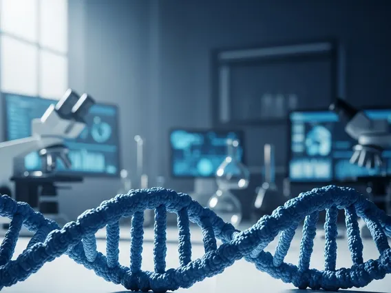
When a diagnosis of testicular cancer is confirmed, the next critical step is to determine what are the stages of testicular cancer? Staging is a standardized process that helps doctors understand if the cancer has spread and, if so, to what extent. This information is vital for guiding treatment decisions and predicting a patient’s outlook. The staging system for testicular cancer is unique compared to many other cancers, primarily due to the significant role of blood tumor markers.
The process of testicular cancer staging explained involves a combination of physical exams, imaging tests (like CT scans of the abdomen, pelvis, and chest), and blood tests to measure specific tumor markers. These markers, including Alpha-fetoprotein (AFP), Human Chorionic Gonadotropin (HCG), and Lactate Dehydrogenase (LDH), are proteins or enzymes that can be elevated in the presence of certain types of testicular cancer. Their levels provide crucial insights into the tumor’s activity and the extent of its spread, complementing the anatomical information from imaging.
The Role of TNM and Tumor Markers
The primary system used for staging most cancers, including testicular cancer, is the TNM system, developed by the American Joint Committee on Cancer (AJCC). TNM stands for:
- T (Tumor): Describes the size and extent of the primary tumor in the testicle.
- N (Nodes): Indicates whether the cancer has spread to nearby lymph nodes and, if so, their size and number.
- M (Metastasis): Refers to whether the cancer has spread to distant parts of the body, such as the lungs, liver, or brain.
For testicular cancer, the TNM classification is further refined by the “S” category, which incorporates serum (blood) tumor marker levels (AFP, HCG, LDH). These markers are categorized into S0 (normal), S1, S2, or S3, based on their elevation levels. This unique integration of biochemical markers makes testicular cancer staging particularly precise, as it reflects not just the physical spread but also the biological activity of the cancer cells.
Clinical vs. Pathological Staging
Understanding how is testicular cancer staged? involves differentiating between clinical and pathological staging. Clinical staging (cTNM) is determined before any surgical removal of the primary tumor. It relies on physical exams, imaging scans, and initial blood tests. This initial staging guides the first steps of treatment, which often involves surgery to remove the affected testicle (orchiectomy).
Pathological staging (pTNM), on the other hand, is determined after the orchiectomy, using a microscopic examination of the removed testicle. This provides more definitive information about the primary tumor (pT). If lymph nodes are also surgically removed (e.g., during a retroperitoneal lymph node dissection, or RPLND), they can be pathologically staged as well (pN). Pathological staging is generally more accurate as it directly assesses the tumor and any involved lymph nodes, offering a clearer picture of the disease’s true extent and helping to fine-tune subsequent treatment plans.
Detailed Breakdown of Testicular Cancer Stages
The classification of testicular cancer stage 1 2 3 provides a clear framework for understanding the progression of the disease. Each stage represents a different extent of cancer spread, influencing both treatment strategies and prognosis.
Stage I: Localized Cancer
Stage I testicular cancer is the earliest form of the disease, indicating that the cancer is confined solely to the testicle. At this stage, there is no evidence of spread to lymph nodes or distant sites. While stages of testicular cancer symptoms typically prompt initial medical consultation, these symptoms (such as a lump or swelling in the testicle) are usually localized. Diagnostic tests confirm that the cancer has not spread beyond the primary tumor. Stage I is often subdivided into IA, IB, and IS, based on the extent of tumor invasion within the testicle and the presence of elevated tumor markers after orchiectomy. For instance, Stage IS indicates that while the primary tumor is removed, tumor markers remain elevated, suggesting microscopic disease that is not yet visible on scans.
Stage II: Regional Spread
Stage II testicular cancer signifies that the disease has spread beyond the testicle to regional lymph nodes, typically those in the retroperitoneum (the area at the back of the abdomen). However, there is no evidence of distant metastasis to other organs. This stage is further categorized into IIA, IIB, and IIC, depending on the size and number of affected lymph nodes. For example, Stage IIA involves smaller lymph nodes, while Stage IIC indicates larger or more numerous affected nodes. The presence of cancer in regional lymph nodes suggests a more aggressive disease course than Stage I, necessitating more intensive treatment approaches.
Stage III: Distant Metastasis
Stage III is the most advanced stage of testicular cancer, characterized by the spread of cancer cells to distant lymph nodes or other organs in the body (distant metastasis). Common sites for metastasis include the lungs, liver, and brain. This stage is also subdivided into IIIA, IIIB, and IIIC, primarily based on the extent of distant spread and the levels of serum tumor markers. For example, Stage IIIA might involve spread to distant lymph nodes or lungs with relatively low tumor marker levels, while Stage IIIC indicates extensive distant spread and/or very high tumor marker levels. The presence of distant metastasis significantly impacts the complexity of treatment and the overall prognosis.
Prognosis and Survival Rates by Stage
The prognosis by testicular cancer stage varies significantly, with earlier detection generally leading to higher survival rates. Testicular cancer is highly curable, even in advanced stages, making it one of the most treatable solid tumors. Survival rates are typically reported as five-year relative survival rates, which compare the survival of cancer patients to that of people in the general population.
According to data from the American Cancer Society and the National Cancer Institute (NCI), the five-year relative survival rates for testicular cancer are:
| Testicular Cancer Stage | Five-Year Relative Survival Rate (Approximate) |
|---|---|
| Stage I (Localized) | 99% |
| Stage II (Regional Spread) | 96% |
| Stage III (Distant Metastasis) | 73% |
| All Stages Combined | 95% |
[Source: American Cancer Society, National Cancer Institute (NCI) SEER Program. These figures are estimates and can vary based on specific cancer type, patient age, overall health, and treatment response.]
It’s important to note that these statistics represent averages and individual outcomes can vary. Factors such as the specific type of germ cell tumor (seminoma vs. non-seminoma), the patient’s overall health, and response to treatment also play a crucial role in prognosis. Even for Stage III disease, advancements in chemotherapy have dramatically improved outcomes, offering significant hope for patients.
Treatment Options Based on Testicular Cancer Stage
Treatment for testicular cancer is highly individualized, with the specific approach largely dictated by the diagnosed testicular cancer stages. The primary goal is to eradicate the cancer while preserving quality of life as much as possible.
- Stage I: For localized cancer, the initial treatment is almost always surgery to remove the affected testicle (radical inguinal orchiectomy). After surgery, patients may undergo active surveillance (close monitoring), a short course of chemotherapy, or radiation therapy, depending on risk factors for recurrence (e.g., presence of lymphovascular invasion).
- Stage II: When cancer has spread to regional lymph nodes, treatment typically involves orchiectomy followed by chemotherapy. In some cases, particularly for non-seminoma, a retroperitoneal lymph node dissection (RPLND) may be performed after chemotherapy or as a primary treatment. Radiation therapy can also be an option for seminoma that has spread to lymph nodes.
- Stage III: For distant metastasis, systemic chemotherapy is the cornerstone of treatment after orchiectomy. The specific chemotherapy regimen depends on the type of testicular cancer (seminoma or non-seminoma) and the risk stratification (good, intermediate, or poor prognosis). Surgery (such as RPLND or resection of lung metastases) may be performed after chemotherapy to remove any remaining cancerous tissue.
The multidisciplinary team, including urologists, oncologists, and radiation oncologists, collaborates to develop the most effective treatment plan, considering the stage, tumor markers, and patient-specific factors.
Life After Staging: Follow-up and Monitoring
After completing initial treatment for any of the testicular cancer stages, ongoing follow-up and monitoring are critical. This surveillance period is designed to detect any signs of recurrence early and to manage potential long-term side effects of treatment. The intensity and duration of follow-up depend on the initial stage of cancer and the treatments received, but it typically lasts for several years.
Regular follow-up usually includes:
- Physical examinations: To check for any new lumps or changes.
- Blood tests: To monitor tumor marker levels (AFP, HCG, LDH), which can indicate recurrence even before it’s visible on scans.
- Imaging scans: Such as CT scans of the abdomen, pelvis, and chest, to check for any returning cancer or new spread. The frequency of these scans decreases over time.
Patients are also educated on the importance of self-examination and reporting any new or concerning symptoms promptly. Long-term monitoring also addresses potential side effects of treatment, such as fertility issues, cardiovascular health, and the risk of secondary cancers, ensuring a holistic approach to patient well-being.
Frequently Asked Questions About Testicular Cancer Stages
How quickly does testicular cancer spread from Stage 1?
The rate at which testicular cancer spreads from Stage 1 varies significantly among individuals and depends on the specific tumor biology. Some cancers may remain localized for an extended period, while others can progress more rapidly. Factors like the presence of lymphovascular invasion (cancer cells in blood or lymphatic vessels) are indicators of a higher risk of spread. Regular follow-up and surveillance after initial treatment are crucial for early detection of any progression, ensuring timely intervention.
Can testicular cancer be cured at Stage 3?
Yes, testicular cancer can often be cured even at Stage 3, which involves distant metastasis. Testicular cancer is highly responsive to chemotherapy, making it one of the most curable solid tumors. While the five-year survival rate for Stage 3 is lower than for earlier stages, it remains remarkably high, often exceeding 70%. Treatment typically involves aggressive systemic chemotherapy, and sometimes surgery to remove any remaining tumors after chemotherapy, leading to successful outcomes for many patients.
What are the main factors determining testicular cancer stage?
The main factors determining testicular cancer stage are the size and extent of the primary tumor (T), whether the cancer has spread to regional lymph nodes (N), and whether it has spread to distant sites or organs (M). Additionally, the levels of specific blood tumor markers (Alpha-fetoprotein, Human Chorionic Gonadotropin, and Lactate Dehydrogenase) play a critical role in refining the stage, particularly for Stage I and Stage III classifications. These factors are combined using the TNM and S (serum marker) systems to assign a definitive stage.