
Slit Lamp Eye Exam
A Slit Lamp Eye Exam is a fundamental diagnostic procedure in ophthalmology, providing a magnified, three-dimensional view of the eye’s structures. This examination is crucial for detecting and monitoring a wide array of ocular conditions, ensuring early intervention and effective management.
Key Takeaways
- A Slit Lamp Eye Exam uses a specialized microscope and a bright light to examine the front and back of the eye.
- Its primary purpose is to detect, diagnose, and monitor various eye conditions affecting different ocular structures.
- The procedure involves the patient resting their chin and forehead while the ophthalmologist views the eye through the slit lamp.
- Conditions such as cataracts, glaucoma, corneal abrasions, and diabetic retinopathy can be diagnosed with this exam.
- It is a painless, non-invasive procedure vital for comprehensive eye health assessment.
What is a Slit Lamp Eye Exam and Its Purpose?
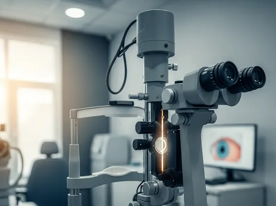
A Slit Lamp Eye Exam is a diagnostic procedure that utilizes a biomicroscope, commonly known as a slit lamp, to provide a highly magnified, three-dimensional view of the eye’s anterior and posterior segments. This instrument combines a powerful light source, which projects a narrow beam (or “slit”) of light, with a stereoscopic microscope, allowing the ophthalmologist to examine the intricate structures of the eye in detail.
The primary purpose of slit lamp examination is to thoroughly assess the health of the eye, identifying abnormalities that might not be visible during a standard eye chart test. It enables clinicians to inspect the eyelids, conjunctiva, cornea, iris, lens, and the front portion of the vitreous humor. With the use of additional lenses, the examination can extend to the retina and optic nerve, providing a comprehensive overview of ocular health. This detailed visualization is essential for the early detection and accurate diagnosis of numerous eye diseases.
How a Slit Lamp Eye Exam is Performed
Performing a Slit Lamp Eye Exam is a straightforward and non-invasive process. The patient is asked to sit comfortably in a chair, placing their chin on a chin rest and their forehead against a support bar. This positioning helps stabilize the head and ensures the eyes are correctly aligned with the slit lamp instrument.
The ophthalmologist then directs a thin, intense beam of light into the patient’s eye, moving it across different areas to illuminate various structures. Simultaneously, they look through the microscope eyepieces to examine the eye’s tissues and fluids. The doctor may ask the patient to look in different directions to allow for a complete view. In some cases, eye drops may be administered to dilate the pupils, which widens them to provide a clearer view of the back of the eye, including the retina and optic nerve. This part of the exam typically takes about 5 to 10 minutes, though it may be longer if pupil dilation is required. While the bright light can be momentarily intense, the procedure itself is painless.
Conditions Diagnosed by Slit Lamp Examination
The detailed view provided by the slit lamp makes it an invaluable tool for diagnosing a wide range of ocular conditions. The conditions diagnosed by slit lamp exam span from common irritations to serious sight-threatening diseases, often allowing for early detection and timely intervention. According to the World Health Organization (WHO), unaddressed eye conditions and vision impairment affect at least 2.2 billion people globally, highlighting the importance of comprehensive eye examinations.
Some of the key conditions that can be identified during this examination include:
- Cataracts: Clouding of the eye’s natural lens.
- Glaucoma: A group of diseases that damage the optic nerve, often due to high intraocular pressure.
- Corneal Abrasions or Ulcers: Scratches or open sores on the cornea.
- Conjunctivitis: Inflammation or infection of the conjunctiva (pink eye).
- Diabetic Retinopathy: Damage to the blood vessels in the retina due to diabetes.
- Macular Degeneration: Deterioration of the macula, the central part of the retina.
- Dry Eye Syndrome: Insufficient lubrication and moisture on the surface of the eye.
- Foreign Bodies: Small particles lodged in the eye.
- Retinal Detachment: Separation of the retina from the underlying tissue (often requires additional lenses for full view).
This comprehensive diagnostic capability underscores the Slit Lamp Eye Exam’s critical role in maintaining eye health and preventing severe vision loss.


