
Sentinel Lymph Node Biopsy
Sentinel Lymph Node Biopsy is a critical diagnostic procedure used in oncology to determine if cancer cells have spread from a primary tumor to the lymphatic system. This technique helps guide treatment decisions and provides important prognostic information for various cancers.
Key Takeaways
- Sentinel Lymph Node Biopsy (SLNB) is a surgical procedure to identify and remove the first lymph node(s) to which cancer cells are most likely to spread.
- It helps determine if cancer has metastasized, influencing treatment plans for cancers like breast cancer and melanoma.
- The procedure involves injecting a tracer (dye or radioactive substance) to locate the sentinel node(s) for removal and pathological examination.
- Recovery typically involves managing mild pain and swelling, with most patients resuming normal activities within a few weeks.
- SLNB can often spare patients from more extensive lymph node dissections, reducing potential side effects.
What is Sentinel Lymph Node Biopsy (SLNB)?
Sentinel Lymph Node Biopsy (SLNB) refers to a surgical procedure performed to identify, remove, and examine the sentinel lymph node(s). The sentinel lymph node is defined as the first lymph node or group of nodes to which cancer cells are most likely to spread from a primary tumor. The primary purpose of SLNB is to determine if cancer has spread beyond its original site, which is crucial for accurate cancer staging and treatment planning.
The sentinel lymph node biopsy meaning is significant in modern oncology because it allows surgeons to assess regional lymph node involvement with minimal invasiveness compared to a complete lymph node dissection. For example, in breast cancer, SLNB has revolutionized staging, as it can prevent unnecessary removal of healthy lymph nodes, thereby reducing the risk of complications like lymphedema. According to the American Cancer Society, SLNB is now a standard procedure for many early-stage breast cancers and melanomas.
The Sentinel Lymph Node Biopsy Procedure
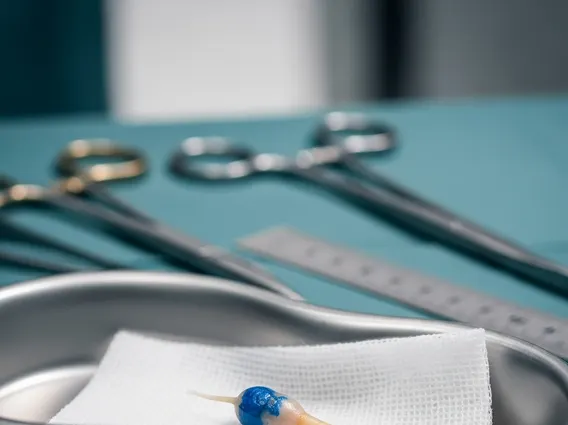
The sentinel lymph node biopsy procedure typically involves two main phases: mapping and surgical removal. Before the surgery, a surgeon or nuclear medicine physician injects a tracer substance near the tumor site. This tracer can be a blue dye, a radioactive substance, or both. The tracer travels through the lymphatic channels, mimicking the path cancer cells would take, and accumulates in the sentinel lymph node(s).
During the surgical phase, the surgeon uses a special device (like a gamma probe for radioactive tracers) or visually identifies the blue-stained nodes to locate the sentinel lymph node(s). Once identified, these nodes are carefully removed through a small incision. The removed nodes are then sent to a pathologist, who examines them under a microscope for the presence of cancer cells. This detailed examination helps determine the extent of cancer spread and guides subsequent treatment decisions.
The steps generally include:
- Tracer Injection: A radioactive tracer or blue dye is injected near the tumor.
- Mapping: The tracer travels to the sentinel lymph node(s), which are then located using a detector or visual inspection.
- Surgical Removal: The identified sentinel node(s) are surgically excised.
- Pathological Analysis: The removed nodes are examined for cancer cells to determine if the cancer has spread.
Sentinel Lymph Node Biopsy Recovery
Sentinel lymph node biopsy recovery generally involves a relatively short period, with most patients returning to their normal activities within a few days to a couple of weeks. Immediately after the procedure, patients may experience some pain, bruising, or swelling at the incision site. If a blue dye was used, the skin around the incision, and sometimes urine, may appear blue or green temporarily. Pain can usually be managed with over-the-counter pain relievers or mild prescription medication.
Patients are typically advised to keep the incision site clean and dry and to avoid strenuous activities or heavy lifting for a period recommended by their surgeon. While SLNB is less invasive than a full lymph node dissection, there is still a small risk of complications such as infection, seroma (fluid collection), or temporary numbness in the area. Long-term complications like lymphedema are significantly less common with SLNB compared to complete lymph node removal. Follow-up appointments are crucial to monitor healing and discuss pathology results, which will inform any further treatment plans.


