Screening Mammogram
A screening mammogram is a vital medical imaging procedure designed for the early detection of breast cancer. This routine examination plays a crucial role in women’s health by identifying potential issues before they become noticeable.
Key Takeaways
- A screening mammogram is an X-ray of the breast used to detect early signs of cancer in individuals without symptoms.
- Early detection through screening significantly improves treatment outcomes and survival rates.
- Recommended frequency varies, typically annually or biennially for women over 40 or 50, depending on risk factors and guidelines.
- The procedure involves brief breast compression and takes only a few minutes, with minimal discomfort.
- Regular screening mammograms are a cornerstone of preventive healthcare for breast cancer.
What is a Screening Mammogram?
A Screening Mammogram is a low-dose X-ray examination of the breast used to look for changes that might indicate early signs of breast cancer in women who do not have any symptoms or noticeable problems. Its primary purpose is to detect breast abnormalities, such as calcifications or masses, that are too small to be felt during a physical exam. By identifying these changes at an early stage, screening mammograms enable timely intervention and potentially less aggressive treatment options.
Unlike a diagnostic mammogram, which is performed when a woman has symptoms like a lump, pain, or nipple discharge, a screening mammogram is a routine preventive measure. It is a quick procedure that typically involves taking two X-ray images of each breast from different angles, providing healthcare providers with a comprehensive view of the breast tissue.
Benefits and Recommended Frequency
The primary benefit of screening mammograms is their ability to detect breast cancer early, often before it has spread. This early detection is critical because it significantly improves the prognosis and survival rates for individuals diagnosed with the disease. For instance, according to the American Cancer Society, regular mammograms have been shown to reduce the risk of dying from breast cancer, particularly for women aged 40 to 74. Early detection can also lead to less extensive surgery and fewer aggressive treatments like chemotherapy.
Guidelines for how often women should get a screening mammogram can vary by age, individual risk factors, and national health organizations. It is always recommended to consult with a healthcare provider to determine the most appropriate schedule for personal health needs. However, general recommendations often include:
- Women aged 40-49: Some organizations recommend starting annual screening, while others suggest discussing with a doctor to weigh benefits and risks.
- Women aged 50-74: Most major health organizations recommend screening every one to two years.
- Women aged 75 and older: Screening recommendations are often individualized based on overall health and life expectancy.
Factors such as a family history of breast cancer, genetic mutations, or a personal history of certain breast conditions may warrant earlier or more frequent screenings. Regular discussions with a physician ensure that screening schedules are tailored to individual risk profiles.
What to Expect During a Screening Mammogram
Knowing what to expect during a screening mammogram can help alleviate any anxiety about the procedure. Upon arrival, you will typically be asked to remove clothing from the waist up and wear a gown. It is important to avoid using deodorants, antiperspirants, lotions, or powders on the day of the exam, as these can appear as abnormalities on the X-ray images.
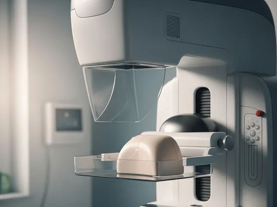
During the mammogram, a technologist will position one breast at a time on a special platform. A clear plastic plate will then gently but firmly press down on the breast for a few seconds. This compression is essential for several reasons: it flattens the breast tissue to allow for a clearer image, spreads out overlapping tissues, reduces the amount of radiation needed, and holds the breast still to prevent blurring. While the compression may cause some discomfort or pressure, it is usually brief. Two images are typically taken of each breast—one from top to bottom and one from side to side. The entire process usually takes about 15 to 20 minutes.
After the images are taken, a radiologist will review them for any suspicious areas. The results are typically communicated to you and your healthcare provider within a few days or weeks. If any areas require further investigation, you may be called back for additional imaging or tests.