
Scintigraphy
Scintigraphy is a sophisticated diagnostic imaging technique within nuclear medicine that provides functional insights into various organs and systems within the body. Unlike traditional imaging methods that focus on anatomical structures, scintigraphy reveals how organs are functioning at a molecular level.
Key Takeaways
- Scintigraphy is a nuclear medicine imaging technique that uses small amounts of radioactive tracers.
- It helps diagnose diseases by showing how organs and tissues function, rather than just their structure.
- The procedure involves injecting a radiotracer, which is then detected by a specialized camera.
- It is a versatile tool used across many medical specialties, including oncology, cardiology, and endocrinology.
- Scintigraphy is non-invasive and provides valuable information for treatment planning and monitoring.
Understanding What Scintigraphy Is
Scintigraphy refers to a diagnostic imaging method that utilizes small, safe amounts of radioactive materials, known as radiotracers, to visualize and assess the function of organs and tissues. This technique is a cornerstone of nuclear medicine, offering a unique perspective on physiological processes that might not be evident with other imaging modalities like X-rays, CT scans, or MRI, which primarily focus on anatomical structures. By observing the distribution and uptake of the radiotracer, clinicians can detect abnormalities in metabolism, blood flow, or cellular activity, providing crucial information for diagnosis and treatment planning.
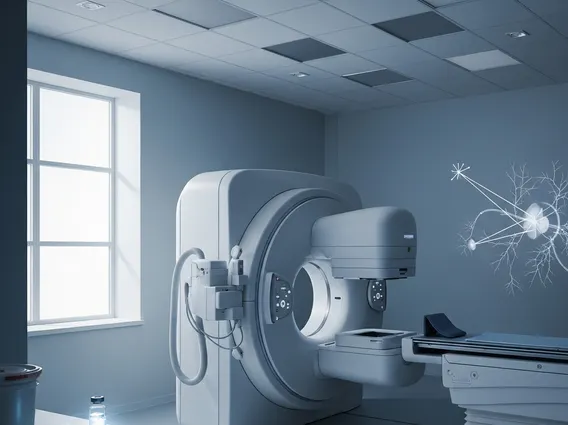
The fundamental principle behind scintigraphy involves the selective accumulation of a radiotracer in specific organs or tissues. Once administered, the radiotracer emits gamma rays, which are then detected by a specialized camera, often called a gamma camera. This camera processes the emissions to create detailed images or 3D reconstructions, illustrating the functional state of the area under investigation. This ability to provide functional information makes scintigraphy invaluable for early disease detection and monitoring disease progression.
The Scintigraphy Procedure: How It Works
The process of how scintigraphy works begins with the administration of a radiotracer into the patient’s body, typically through intravenous injection, but sometimes orally or by inhalation, depending on the specific scan. These radiotracers are designed to target particular organs, cells, or metabolic pathways. For instance, a bone scan might use a radiotracer that is absorbed by bone-forming cells, while a cardiac scan might use one that highlights blood flow to the heart muscle.
Once inside the body, the radiotracer travels and accumulates in the target area. The patient then lies on a table while a gamma camera moves around or over the body, detecting the gamma rays emitted by the radiotracer. The camera converts these emissions into electrical signals, which a computer then processes to create images. These images show “hot spots” where the tracer accumulates more (indicating increased activity or blood flow) or “cold spots” where it accumulates less (suggesting reduced activity or blockages). The duration of the scan can vary from minutes to several hours, depending on the type of study and how quickly the tracer distributes and clears from the body.
Applications and Varieties of Scintigraphy Scans
The scintigraphy uses and benefits are extensive, spanning numerous medical specialties due to its ability to provide functional insights. One of the primary benefits is its sensitivity in detecting diseases at early stages, often before structural changes become apparent on other imaging tests. This can lead to earlier intervention and improved patient outcomes. For example, in oncology, scintigraphy can identify metastatic cancer spread to bones long before it causes pain or visible changes on X-rays. According to the World Nuclear Association, more than 40 million nuclear medicine procedures, including various forms of scintigraphy, are performed each year globally, highlighting their widespread utility and importance in modern medicine.
There are many types of scintigraphy scans, each tailored to assess specific organs or conditions. These include:
- Bone Scans: Used to detect fractures, infections, tumors, and bone metastases.
- Cardiac Scans (Myocardial Perfusion Scans): Evaluate blood flow to the heart muscle, helping diagnose coronary artery disease and assess heart function.
- Thyroid Scans: Assess thyroid gland function and identify nodules or hyperthyroidism.
- Kidney Scans (Renal Scans): Evaluate kidney function, blood flow, and identify obstructions.
- Gallium Scans: Detect inflammation, infection, and certain types of cancer.
- Lung Scans (V/Q Scans): Used to diagnose pulmonary embolism by evaluating ventilation and perfusion in the lungs.
- Brain Scans (SPECT Scans): Can assess blood flow to the brain, aiding in the diagnosis of stroke, dementia, and seizures.
These diverse applications underscore scintigraphy’s role as a versatile and indispensable diagnostic tool, offering unique functional information that complements other imaging techniques to provide a comprehensive view of a patient’s health.


