
Microscope
A Microscope is an essential scientific instrument that magnifies small objects, enabling detailed observation of structures invisible to the naked eye. It plays a pivotal role across various scientific disciplines, particularly in medicine, clinical diagnostics, and oncology.
Key Takeaways
- A microscope is an optical instrument that magnifies minute objects, revealing their intricate details.
- Microscopes operate by using lenses or electron beams to magnify samples, enhancing resolution and clarity.
- They are indispensable in medical diagnostics for identifying pathogens, analyzing tissue samples, and detecting cellular abnormalities.
- The evolution of microscopy, from early optical devices to advanced electron microscopes, has revolutionized our understanding of biology and disease.
- Different types of microscopes are employed based on the required magnification, resolution, and sample characteristics.
What is a Microscope?
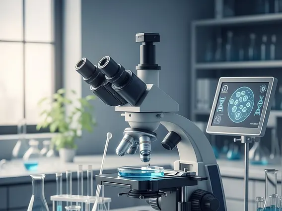
A Microscope refers to an instrument designed to produce magnified images of small objects, allowing for the examination of fine details and structures that are too small to be seen with the unaided eye. In medical and clinical contexts, microscopes are fundamental tools for diagnosing diseases, studying cellular processes, and identifying microorganisms. Their ability to reveal the microscopic world has profoundly impacted pathology, microbiology, hematology, and oncology, providing critical insights into the nature of health and disease.
The core function of a microscope is to increase both magnification and resolution. Magnification refers to the enlargement of an object’s image, while resolution is the ability to distinguish between two closely spaced points. High resolution is crucial in clinical settings for accurately identifying cellular abnormalities or distinguishing between different types of bacteria, directly influencing diagnostic accuracy and treatment decisions.
How Microscopes Work and Their Diverse Applications
Microscopes operate on principles that vary depending on their type, but the fundamental goal is to magnify and resolve fine details of a sample. Optical microscopes, for instance, use a system of lenses to focus light through or reflected from a specimen, creating a magnified image that can be viewed directly or captured digitally. The interplay between the objective lens (close to the specimen) and the eyepiece lens (where the observer looks) determines the total magnification and the clarity of the image.
The applications of a microscope are vast and critical in clinical practice. What is a microscope used for? In medicine, microscopes are indispensable for:
- Pathology: Examining tissue biopsies and surgical specimens to diagnose cancer, inflammatory conditions, and other diseases.
- Microbiology: Identifying bacteria, fungi, parasites, and viruses in patient samples to diagnose infectious diseases. The World Health Organization (WHO) emphasizes microscopic examination as a cornerstone for diagnosing many infectious diseases, such as tuberculosis and malaria, which affect millions globally.
- Hematology: Analyzing blood smears to detect blood disorders like anemia, leukemia, and parasitic infections.
- Cytology: Screening for abnormal cells, such as in Pap tests for cervical cancer detection.
How does a microscope work? Beyond basic light transmission, advanced microscopes employ various techniques, including phase contrast for unstained living cells, fluorescence for specific molecular tagging, and electron beams for ultra-high resolution imaging of cellular ultrastructure. These methods allow clinicians and researchers to visualize different aspects of biological samples, from whole cells to subcellular organelles and macromolecules.
Evolution and Types of Microscopes
The History of the microscope dates back to the late 16th and early 17th centuries, with early pioneers like Zacharias Janssen and later Antonie van Leeuwenhoek and Robert Hooke making significant advancements. Leeuwenhoek is often credited with developing simple microscopes that achieved remarkable magnification for his time, allowing him to observe bacteria and other microorganisms. Hooke’s compound microscope, detailed in his 1665 work “Micrographia,” provided some of the earliest detailed drawings of microscopic life, including plant cells.
Over centuries, microscopy has evolved dramatically, leading to a diverse array of instruments. Types of microscopes and their uses vary widely based on their underlying technology and specific applications:
- Compound Light Microscope: The most common type, used for viewing stained or unstained cells and tissues at magnifications up to 1000x. It is a staple in clinical laboratories for routine diagnostics.
- Stereo Microscope (Dissecting Microscope): Provides a 3D view of larger specimens at lower magnifications, useful for dissecting and examining whole organisms or larger tissue samples.
- Electron Microscopes (TEM and SEM): Utilize beams of electrons instead of light to achieve much higher magnification (up to millions of times) and resolution. Transmission Electron Microscopes (TEM) are used to view internal structures, while Scanning Electron Microscopes (SEM) provide detailed surface topography. These are crucial for studying viruses, cellular organelles, and material science.
- Fluorescence Microscope: Employs fluorescent dyes to highlight specific molecules or structures within a cell, vital for molecular biology and immunology research and diagnostics.
Each type offers unique advantages, contributing to a comprehensive understanding of biological systems at different scales, from macroscopic observations to atomic-level detail, thereby continually advancing medical science and clinical care.


