Melanoma Stages
Understanding melanoma stages is crucial for both patients and healthcare providers, as it directly influences treatment decisions and prognosis. This article provides a comprehensive overview of how melanoma, a serious form of skin cancer, is staged, from its earliest forms to advanced disease.
Key Takeaways
- Melanoma staging uses the TNM system (Tumor, Node, Metastasis) to classify the extent of the cancer.
- Early stages (0, I, II) indicate localized disease, with prognosis largely dependent on tumor thickness and ulceration.
- Advanced stages (III, IV) involve spread to regional lymph nodes or distant organs, requiring more aggressive treatment strategies.
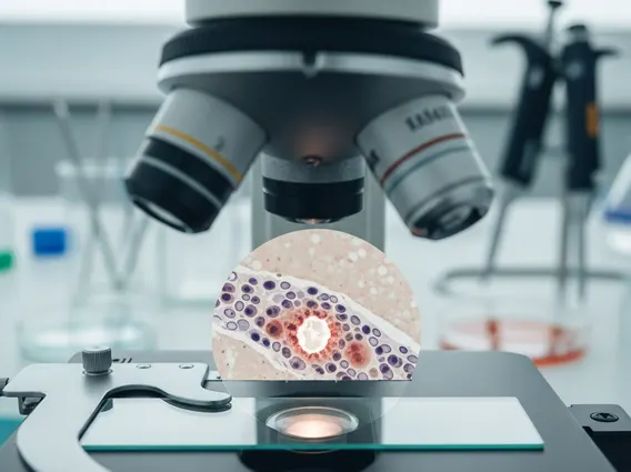
- Staging is determined through biopsy, pathological examination, and imaging, including sentinel lymph node biopsy.
- Early detection and accurate staging are critical for effective treatment and improving long-term survival rates.
What Are the Stages of Melanoma?
The process of determining what are the stages of melanoma involves a detailed assessment of the cancer’s characteristics and spread. This system, known as the TNM staging system developed by the American Joint Committee on Cancer (AJCC), categorizes melanoma into stages 0 through IV, with higher numbers indicating more extensive disease. Each stage provides vital information about the tumor’s size, whether it has spread to nearby lymph nodes, and if it has metastasized to distant parts of the body. This comprehensive approach ensures that melanoma staging explained is clear and actionable for medical professionals.
Why Staging is Crucial
Accurate staging is paramount because it dictates the most appropriate treatment plan and offers insights into a patient’s prognosis. For instance, early-stage melanomas are often curable with surgery alone, while advanced stages may require a combination of therapies such as immunotherapy, targeted therapy, or radiation. Staging also allows for a better understanding melanoma progression, helping doctors anticipate potential challenges and tailor follow-up care. According to the National Cancer Institute, the 5-year survival rate for localized melanoma is 99%, emphasizing the importance of early detection and precise staging.
Key Factors in Staging
Several key factors contribute to determining the stage of melanoma:
- Tumor Thickness (Breslow Depth): This is the most important factor, measuring how deep the melanoma has grown into the skin. Thicker tumors generally indicate a higher risk of spread.
- Ulceration: The presence of an open sore or break in the skin over the melanoma suggests a more aggressive tumor.
- Mitotic Rate: The number of dividing cancer cells observed under a microscope, which can indicate how quickly the tumor is growing.
- Lymph Node Involvement: Whether cancer cells have spread to nearby lymph nodes.
- Distant Metastasis: If the cancer has spread to distant organs or lymph nodes far from the primary tumor.
- Lactate Dehydrogenase (LDH) Levels: Elevated levels of this enzyme in the blood can indicate more advanced disease, particularly in Stage IV melanoma.
Early Stage Melanoma: 0, I, and II
The initial stages of melanoma represent localized disease, meaning the cancer is confined to the skin and has not spread to lymph nodes or distant sites. Recognizing these different stages of melanoma skin cancer early significantly improves treatment outcomes.
Stage 0: Melanoma In Situ
Stage 0 melanoma, also known as melanoma in situ, is the earliest form of the disease. In this stage, the abnormal melanocytes (pigment-producing cells) are found only in the outermost layer of the skin, the epidermis, and have not invaded deeper tissues. Because it is non-invasive, melanoma in situ is highly curable, typically with surgical removal alone. There are generally no specific melanoma stage 0 to 4 symptoms unique to Stage 0 beyond the visible lesion itself, which might be an unusual mole.
Stages I and II: Localized Disease
Stages I and II melanoma are characterized by invasive tumors that have grown deeper into the skin but have not yet spread to nearby lymph nodes or distant sites. The primary distinction between these stages lies in the tumor’s thickness and the presence or absence of ulceration:
- Stage I: Melanomas are relatively thin (up to 2.0 mm thick) and may or may not be ulcerated. The risk of spread is low, and surgical excision is the standard treatment.
- Stage II: These melanomas are thicker (greater than 2.0 mm, up to 4.0 mm or more) and/or have ulceration. While still localized, Stage II carries a higher risk of recurrence and potential spread compared to Stage I, prompting more aggressive surgical margins and close monitoring.
For both Stage I and II, the primary goal is complete surgical removal of the tumor with clear margins, often followed by regular surveillance.
Advanced Melanoma: Stages III and IV
When melanoma progresses beyond the primary tumor site, it is classified as advanced. Understanding the distinction between early vs advanced melanoma stages is critical for managing the disease effectively.
Stage III: Regional Spread
Stage III melanoma indicates that the cancer has spread from the primary tumor to nearby lymph nodes or to other skin areas or lymphatic channels near the primary tumor (known as satellite or in-transit metastases). Even if the primary tumor was small, the presence of cancer cells in the lymph nodes significantly increases the risk of further spread. The extent of lymph node involvement (number of affected nodes, size of metastases) further subdivides Stage III. At this stage, patients might experience enlarged lymph nodes, or new skin lesions appearing between the primary tumor site and the regional lymph nodes. Treatment often involves surgical removal of the affected lymph nodes (lymphadenectomy), potentially followed by adjuvant therapies like immunotherapy or targeted therapy to reduce the risk of recurrence.
Stage IV: Distant Metastasis
Stage IV melanoma is the most advanced stage, meaning the cancer has spread to distant organs or lymph nodes far from the original tumor site. Common sites for distant metastasis include the lungs, liver, brain, bones, and other parts of the skin. At this stage, melanoma stage 0 to 4 symptoms can become more systemic and varied, depending on the organs affected. For example, lung metastases might cause shortness of breath, brain metastases could lead to headaches or neurological changes, and liver metastases might cause abdominal pain or jaundice. While Stage IV melanoma is challenging to treat, significant advancements in immunotherapy and targeted therapies have improved outcomes for many patients, offering hope where options were once limited. Treatment focuses on controlling the disease, managing symptoms, and improving quality of life.
How Melanoma Staging is Determined
Accurate staging is a multi-step process involving various diagnostic procedures. This section details how is melanoma staged, from initial biopsy to comprehensive imaging.
Biopsy and Pathology
The first and most critical step in staging melanoma is the biopsy of the suspicious lesion. A dermatologist or surgeon removes a sample of the tissue, which is then sent to a pathologist for microscopic examination. The pathologist determines if cancer cells are present and, if so, identifies key characteristics:
- Breslow Thickness: The depth of the tumor invasion, measured in millimeters.
- Ulceration: Presence or absence of an open sore on the tumor surface.
- Mitotic Rate: The number of dividing cancer cells per square millimeter, indicating growth speed.
- Clark Level: Describes the level of invasion into the skin layers (less commonly used than Breslow thickness but still provides context).
These pathological findings are fundamental to determining the initial T (Tumor) category of the TNM system.
Imaging and Lymph Node Evaluation
Once the primary tumor characteristics are known, further tests may be conducted to assess for regional or distant spread:
- Sentinel Lymph Node Biopsy (SLNB): For melanomas of a certain thickness (typically >0.8 mm or with ulceration), an SLNB may be performed. A radioactive tracer and/or blue dye is injected near the melanoma to identify the first lymph node(s) to which cancer cells would likely spread (the sentinel node). These nodes are then surgically removed and examined for cancer cells. If cancer is found, it indicates regional spread (N category).
- Imaging Scans: For higher-risk Stage II, Stage III, and all Stage IV melanomas, imaging tests are used to look for distant spread (M category). These may include:
- Computed Tomography (CT) scans: To visualize internal organs and lymph nodes.
- Positron Emission Tomography (PET) scans: To detect metabolically active cancer cells throughout the body.
- Magnetic Resonance Imaging (MRI) scans: Particularly useful for detecting brain metastases.
- Blood Tests: Blood tests, including lactate dehydrogenase (LDH) levels, can provide additional prognostic information, especially in advanced stages.
The combination of these findings allows oncologists to precisely classify the melanoma stages and develop an individualized treatment plan.
Prognosis and Treatment by Stage
The prognosis for melanoma varies significantly depending on its stage at diagnosis, highlighting the critical role of early detection. Generally, the earlier the stage, the better the prognosis and the higher the chance of a cure. Treatment strategies are tailored to the specific stage, aiming to remove the cancer, prevent recurrence, and manage symptoms.
For Stage 0, I, and II melanoma, the primary treatment is surgical excision of the tumor with a margin of healthy tissue. For Stage II, wider margins may be taken, and an SLNB might be performed to check for microscopic spread to lymph nodes. The 5-year survival rate for localized melanoma (Stages I and II) is approximately 99%, according to the American Cancer Society, underscoring the effectiveness of early intervention.
Stage III melanoma, which involves regional lymph node spread, often requires more aggressive treatment. This typically includes surgical removal of the affected lymph nodes (lymphadenectomy). Following surgery, adjuvant therapies such as immunotherapy (e.g., PD-1 inhibitors) or targeted therapy (for melanomas with specific genetic mutations like BRAF) may be recommended to reduce the risk of recurrence. Radiation therapy might also be used in certain situations. The 5-year survival rate for regional melanoma (Stage III) is around 71%.
Stage IV melanoma, characterized by distant metastasis, is the most challenging to treat. The focus shifts to systemic therapies aimed at controlling the disease throughout the body, managing symptoms, and improving quality of life. Treatment options include:
- Immunotherapy: Drugs that boost the body’s immune system to fight cancer cells, such as checkpoint inhibitors.
- Targeted Therapy: Medications that target specific genetic mutations within the cancer cells (e.g., BRAF and MEK inhibitors for BRAF-mutated melanoma).
- Chemotherapy: Traditional anti-cancer drugs, though less commonly used as a primary treatment for melanoma today due to the efficacy of newer therapies.
- Radiation Therapy: Used to shrink tumors, relieve pain, or treat metastases in specific areas like the brain or bones.
- Surgery: May be used to remove isolated metastases or manage complications.
While the prognosis for Stage IV melanoma is generally poorer, with a 5-year survival rate of about 32%, advancements in immunotherapy and targeted therapies have significantly improved outcomes for many patients in recent years. Continued research offers ongoing hope for new and more effective treatments across all melanoma stages.
Frequently Asked Questions
What is the most important factor in melanoma staging?
The most critical factor in melanoma staging is the Breslow depth, which measures the tumor’s thickness in millimeters. This depth directly correlates with the risk of the cancer spreading. Thicker tumors indicate a higher likelihood of invasion into deeper skin layers and potential metastasis, making it a primary determinant in classifying melanoma into its various stages and guiding subsequent treatment decisions.
Can melanoma stage change over time?
Yes, melanoma stage can change over time. If a melanoma initially diagnosed at an early stage recurs or spreads to lymph nodes or distant organs, its stage will be reclassified to reflect the progression of the disease. Regular follow-up and surveillance are essential for detecting any changes, allowing for timely adjustments to the treatment plan and continued management of the cancer.
What are the general survival rates for early vs. advanced melanoma?
Survival rates vary significantly between early and advanced melanoma. For localized melanoma (Stages 0, I, and II), the 5-year survival rate is very high, often exceeding 90%. However, for advanced melanoma (Stages III and IV), where the cancer has spread to regional lymph nodes or distant organs, the 5-year survival rates decrease. Early detection and treatment are therefore paramount for improving long-term outcomes.