
Mammotome
Mammotome refers to an advanced, minimally invasive biopsy system used primarily for the diagnosis of breast abnormalities. This technology plays a crucial role in the early detection and characterization of suspicious breast lesions, offering a less invasive alternative to traditional surgical biopsies.
Key Takeaways
- Mammotome is a vacuum-assisted biopsy (VAB) system designed for precise tissue sampling.
- It is primarily utilized in breast diagnostics to investigate suspicious lesions identified through imaging.
- The procedure is image-guided, ensuring accurate targeting and collection of tissue samples.
- It offers significant advantages over traditional surgical biopsies, including minimal invasiveness and reduced recovery time.
- Mammotome technology contributes to definitive diagnoses, aiding in timely and appropriate patient management.
What is Mammotome?
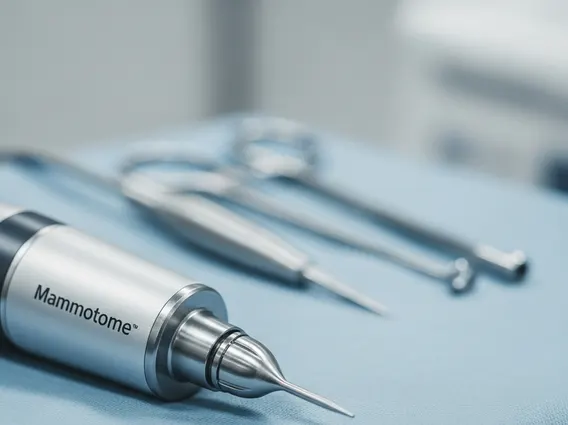
Mammotome is a sophisticated vacuum-assisted biopsy (VAB) system designed to accurately collect tissue samples from suspicious areas within the breast. This technology is instrumental in the diagnostic process for breast cancer and other breast conditions, particularly for lesions that are non-palpable or difficult to access, often detected through imaging modalities like mammography, ultrasound, or magnetic resonance imaging (MRI). Mammotome is used for the definitive diagnosis of breast abnormalities, helping to determine whether a lesion is benign (non-cancerous), malignant (cancerous), or atypical. By providing sufficient tissue for pathological examination, it facilitates a precise diagnosis, which is critical for guiding subsequent treatment decisions. Early detection through methods like biopsy significantly improves breast cancer outcomes, as highlighted by health organizations such as the American Cancer Society.
Mammotome Breast Biopsy Procedure Explained
The Mammotome breast biopsy procedure explained involves several key steps, all performed with precision and patient comfort in mind. The procedure typically begins with local anesthesia to numb the breast area, ensuring the patient experiences minimal discomfort. Image guidance, such as ultrasound, stereotactic mammography, or MRI, is then used to precisely locate the suspicious lesion.
Once the lesion is accurately identified, a small incision, usually about 3-4 mm, is made in the skin. A specialized probe is then inserted through this incision and advanced to the target area. The Mammotome system works by utilizing a vacuum to gently draw tissue into the probe’s opening. A rotating blade within the probe then cuts and collects the tissue samples, which are then transported out of the breast through the same probe without needing to reinsert or withdraw the probe multiple times. This process allows for the collection of multiple samples from various parts of the lesion, ensuring comprehensive tissue analysis. After sufficient samples are collected, the probe is removed, and a small marker clip may be placed at the biopsy site to facilitate future follow-up if needed. The tiny incision is then closed, usually with a sterile strip, requiring no stitches. The entire process typically takes less than an hour, and patients can usually resume normal activities shortly thereafter, with minimal recovery time.
Benefits of Mammotome Technology
The Mammotome technology benefits are numerous, offering significant advantages over traditional open surgical biopsies for breast lesion diagnosis. One of the primary benefits is its minimally invasive nature. Unlike surgical biopsies that require a larger incision and may leave a noticeable scar, Mammotome uses a very small incision, resulting in less scarring and a better cosmetic outcome. This also translates to reduced pain and a quicker recovery period for the patient.
Furthermore, Mammotome offers exceptional accuracy in tissue sampling. Its image-guided approach ensures that samples are taken directly from the suspicious area, reducing the chance of sampling error. The vacuum-assisted mechanism allows for the collection of larger and more numerous tissue samples through a single insertion, providing pathologists with more comprehensive material for a definitive diagnosis. This can often eliminate the need for a subsequent surgical biopsy if the initial results are conclusive.
Here are some key advantages:
- Minimally Invasive: Small incision, less scarring, and reduced discomfort compared to surgical biopsy.
- High Accuracy: Image guidance ensures precise targeting of lesions, even non-palpable ones.
- Comprehensive Sampling: Ability to collect multiple, larger tissue samples through a single insertion.
- Faster Recovery: Patients typically experience less downtime and can return to normal activities quickly.
- Outpatient Procedure: Usually performed in an outpatient setting under local anesthesia.
- Reduced Risk: Lower risk of complications such as infection or hematoma compared to surgery.
These benefits collectively make Mammotome a preferred method for diagnosing breast abnormalities, contributing to more efficient and patient-friendly diagnostic pathways in breast health.


