Glioneuronal Tumor
A glioneuronal tumor is a type of central nervous system neoplasm characterized by a mixture of both glial and neuronal cells. These tumors exhibit diverse clinical behaviors and pathological features, making accurate diagnosis and tailored treatment crucial.

Key Takeaways
- Glioneuronal Tumor is a rare brain or spinal cord tumor comprising both glial and neuronal cells.
- Symptoms vary widely depending on the tumor’s location, with seizures being a common presentation.
- Diagnosis typically involves advanced imaging like MRI, followed by a definitive biopsy and histopathological analysis.
- Treatment primarily focuses on surgical removal, with radiation or chemotherapy considered for specific cases.
- Prognosis is generally favorable for low-grade tumors, but long-term monitoring is essential.
What is a Glioneuronal Tumor?
A Glioneuronal Tumor refers to a heterogeneous group of central nervous system (CNS) neoplasms that contain both glial and neuronal components. These tumors are relatively rare, accounting for a small percentage of all brain and spinal cord tumors. They can arise in various locations within the CNS, including the cerebral hemispheres, cerebellum, brainstem, and spinal cord, with their specific characteristics often influenced by their origin.
These tumors are typically slow-growing and often classified as low-grade, though some variants can exhibit more aggressive behavior. The World Health Organization (WHO) classification of CNS tumors recognizes several distinct types of glioneuronal tumors, each with unique pathological features and clinical implications. Understanding the specific subtype is critical for determining prognosis and guiding therapeutic strategies.
Glioneuronal Tumor Symptoms, Signs, and Diagnosis
The Glioneuronal tumor symptoms and signs are highly variable and depend significantly on the tumor’s size, location, and growth rate within the brain or spinal cord. Common presentations often include neurological symptoms such as seizures, which are particularly prevalent in supratentorial tumors. Other signs may involve persistent headaches, nausea, vomiting, and focal neurological deficits like weakness in limbs, vision changes, or difficulties with speech and coordination. In children, developmental delays or changes in behavior might also be observed.
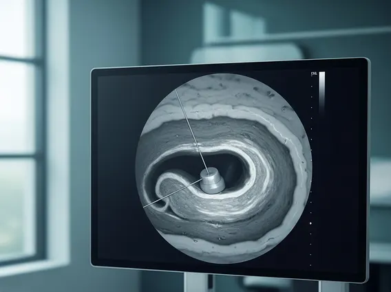
Diagnosing glioneuronal tumors typically begins with a thorough neurological examination and a review of the patient’s medical history. Imaging studies are crucial for initial detection and characterization. Magnetic Resonance Imaging (MRI) with contrast is the preferred method, as it provides detailed images of brain structures and can help identify the tumor’s exact location, size, and relationship to surrounding tissues. While imaging can suggest the presence of a glioneuronal tumor, a definitive diagnosis requires a biopsy.
During a biopsy, a small sample of the tumor tissue is surgically removed and then examined under a microscope by a neuropathologist. This histopathological analysis confirms the presence of both glial and neuronal elements and helps classify the specific subtype of glioneuronal tumor. Molecular testing may also be performed on the tissue sample to identify specific genetic mutations or biomarkers, which can further refine the diagnosis and inform treatment decisions. For instance, some glioneuronal tumors are associated with specific BRAF mutations, which can be targeted by certain therapies.
Glioneuronal Tumor Treatment Options
The primary goal of Glioneuronal tumor treatment options is to remove as much of the tumor as safely possible, alleviate symptoms, and prevent recurrence. The choice of treatment strategy is highly individualized, taking into account the tumor’s type, location, size, the patient’s age, overall health, and the presence of any specific genetic markers.
The most common and often most effective treatment is surgical resection. If the tumor is accessible and can be removed without causing significant neurological damage, complete surgical removal (gross total resection) is the preferred approach. This can often lead to a cure, especially for low-grade tumors. For tumors that cannot be fully removed due to their location or proximity to critical brain structures, a partial resection may be performed to reduce tumor bulk and alleviate symptoms.
Other treatment modalities may be considered, either as adjuvant therapy after surgery or as primary treatment for unresectable tumors:
- Radiation Therapy: This may be used for residual tumor after surgery, for tumors that are higher grade, or for those that recur. It uses high-energy rays to kill cancer cells or slow their growth.
- Chemotherapy: While less common for many low-grade glioneuronal tumors, chemotherapy may be considered for more aggressive subtypes, recurrent tumors, or when other treatments are not feasible.
- Targeted Therapy: For tumors with specific molecular alterations, such as BRAF V600E mutations, targeted therapies that block these specific pathways may be an option. These treatments are often more precise and can have fewer side effects than traditional chemotherapy.
- Observation: In some cases, particularly for very small, asymptomatic, and slow-growing tumors, a “watch and wait” approach with regular MRI scans may be adopted, especially if the risks of intervention outweigh the benefits.
A multidisciplinary team, including neurosurgeons, neuro-oncologists, radiation oncologists, and neuropathologists, typically collaborates to develop the most appropriate treatment plan and provide comprehensive care for patients with glioneuronal tumors.