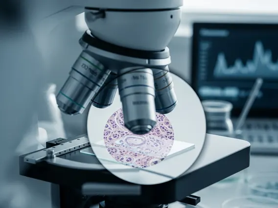
Fusion Biopsy
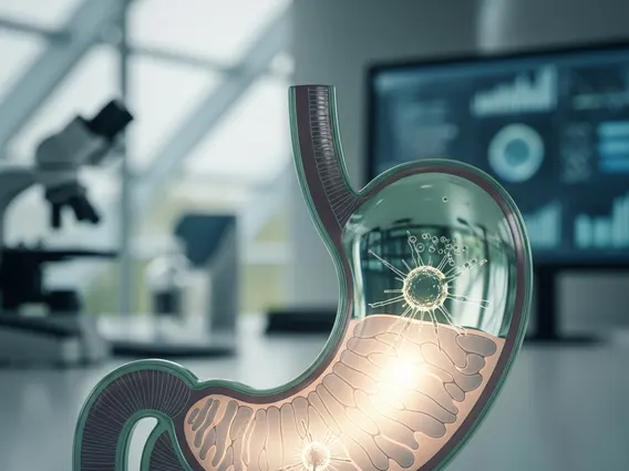
Fusion Biopsy is an advanced diagnostic technique that combines real-time imaging with pre-existing high-resolution scans to guide tissue sampling with greater precision. This method significantly enhances the accuracy of detecting abnormal tissues, particularly in oncology.
Key Takeaways
- Fusion Biopsy integrates MRI and ultrasound images to create a detailed, real-time map for targeted tissue sampling.
- It offers enhanced accuracy compared to traditional, untargeted biopsy methods.
- The procedure is particularly valuable in the diagnosis of prostate cancer, helping to identify clinically significant lesions.
- By precisely targeting suspicious areas, Fusion Biopsy can reduce the number of samples needed and improve diagnostic yield.
What is Fusion Biopsy?
Fusion Biopsy refers to a sophisticated medical procedure that merges images from different diagnostic modalities to create a more comprehensive and accurate guide for tissue sampling. This technique typically combines magnetic resonance imaging (MRI) scans, which provide detailed anatomical and pathological information, with real-time ultrasound imaging. The fusion of these images allows clinicians to visualize suspicious areas identified on the MRI with greater clarity during the biopsy procedure, leading to more precise targeting of abnormal tissue.
This advanced approach addresses limitations of traditional biopsies, which often rely solely on real-time ultrasound that may not clearly distinguish all suspicious lesions. By overlaying the MRI data onto the live ultrasound feed, medical professionals can accurately navigate to and sample specific areas of concern, improving the diagnostic yield and reducing the need for repeat procedures. This precision is crucial for conditions where early and accurate diagnosis significantly impacts treatment outcomes.
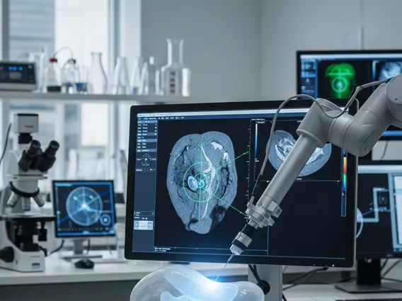
MRI-Ultrasound Fusion Biopsy Procedure Explained
The MRI-Ultrasound Fusion Biopsy procedure involves several key steps designed to maximize accuracy and minimize discomfort. It begins with a pre-biopsy MRI scan, which identifies and maps any suspicious lesions within the target organ, such as the prostate. These MRI images are then digitally fused with real-time ultrasound images during the biopsy itself, creating a dynamic, three-dimensional map that guides the biopsy needle directly to the areas of concern.
During the procedure, the patient is positioned, and local anesthesia is administered to ensure comfort. The ultrasound probe is inserted, and its live feed is synchronized with the previously acquired MRI data. Specialized software overlays the MRI-identified targets onto the live ultrasound screen. This integrated view allows the physician to precisely steer the biopsy needle to the exact location of the suspicious lesion, ensuring that tissue samples are collected from the most relevant areas. The samples are then sent to a pathology lab for analysis. The typical steps include:
- Pre-biopsy MRI: High-resolution MRI scans are performed to identify and characterize suspicious lesions.
- Image Fusion: MRI data is digitally merged with real-time ultrasound images using specialized software.
- Targeted Biopsy: The fused images guide the physician in precisely positioning the biopsy needle to sample the identified lesions.
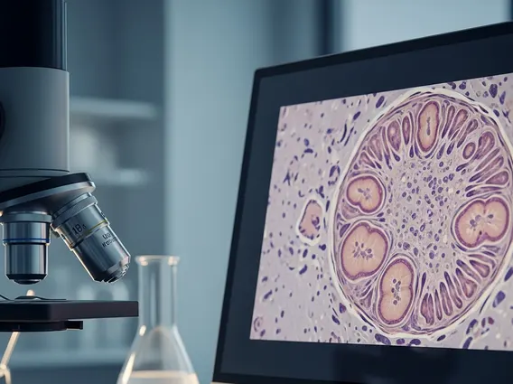
- Pathological Analysis: Collected tissue samples are examined under a microscope to confirm or rule out disease.
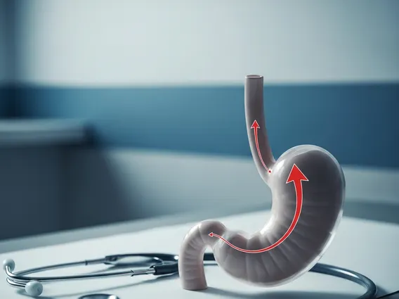
Fusion Biopsy for Prostate Cancer Diagnosis
The application of Fusion Biopsy for prostate cancer diagnosis has revolutionized the detection of this common malignancy. Prostate cancer often presents as subtle lesions that may be difficult to visualize with standard ultrasound alone. MRI, however, excels at identifying and characterizing these lesions, particularly those that are clinically significant and require treatment. By combining the strengths of both imaging modalities, Fusion Biopsy significantly improves the ability to detect aggressive prostate cancers that might otherwise be missed.
Studies have shown that Fusion Biopsy can detect more clinically significant prostate cancers compared to traditional systematic biopsies, which involve taking random samples from across the prostate. For instance, a meta-analysis published in the European Urology journal indicated that MRI-targeted biopsies, including fusion biopsies, detected significantly more clinically significant prostate cancers and fewer insignificant cancers compared to systematic biopsies. This targeted approach not only enhances diagnostic accuracy but also helps to reduce the over-diagnosis of low-risk cancers, potentially preventing unnecessary treatments and their associated side effects. This precision allows for more informed treatment decisions, tailoring interventions to the specific needs of each patient.