Fna Biopsy
An FNA biopsy is a common diagnostic procedure used to investigate lumps or masses in various parts of the body. It offers a minimally invasive way to collect cells for examination, aiding in the diagnosis of conditions ranging from benign cysts to malignant tumors.
Key Takeaways
- FNA biopsy is a minimally invasive procedure that collects cell samples for diagnostic purposes.
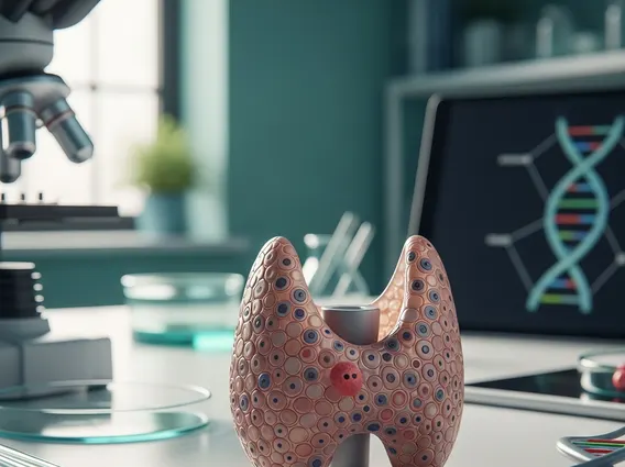
- It is commonly used to evaluate suspicious lumps in areas like the thyroid, breast, or lymph nodes.
- The procedure involves inserting a thin needle to aspirate cells, often guided by imaging.
- Results can classify findings as benign, malignant, or indeterminate, guiding further medical steps.
- While generally safe, potential risks of FNA biopsy include minor bruising, swelling, or infection.
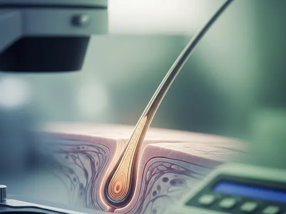
What is an FNA biopsy (Fine Needle Aspiration)?
FNA biopsy, or Fine Needle Aspiration biopsy, is a diagnostic procedure used to investigate suspicious lumps or masses. It involves using a very thin, hollow needle to extract a small sample of cells or fluid from the abnormal area. This sample is then sent to a pathology lab for microscopic examination by a pathologist. The primary goal of an FNA biopsy is to determine if a lump is benign (non-cancerous) or malignant (cancerous), or to identify other specific conditions. It is a quick, relatively non-invasive, and often outpatient procedure, making it a preferred initial diagnostic tool for many types of masses, including those found in the thyroid, breast, lymph nodes, salivary glands, and soft tissues. This procedure provides crucial information that helps clinicians decide on the next steps in a patient’s care, whether that involves further testing, monitoring, or treatment.
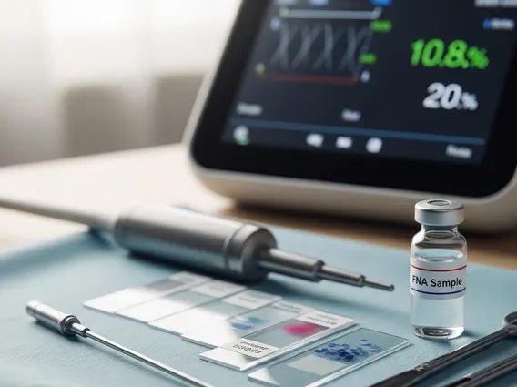
How is an FNA biopsy performed?
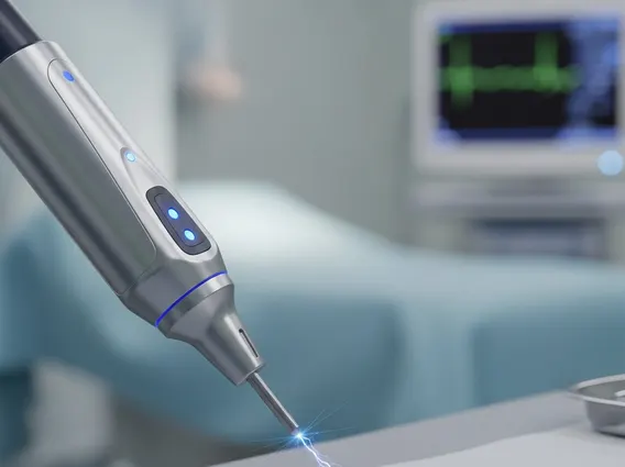
The process of an FNA biopsy typically begins with the patient positioned comfortably. The area of the lump is cleaned with an antiseptic solution. In some cases, a local anesthetic may be administered to numb the skin, though this is not always necessary due to the thinness of the needle. For palpable lumps (those that can be felt), the physician will guide the needle into the mass by touch. For deeper or non-palpable lesions, imaging guidance, such as ultrasound or CT scan, is often used to ensure precise needle placement. Once the needle is in the target area, the physician applies suction using a syringe to draw cells and fluid into the needle. The needle may be moved back and forth gently several times to collect an adequate sample. This aspiration process usually takes only a few seconds. After the sample is collected, the needle is withdrawn, and pressure is applied to the site to minimize bleeding. The collected cells are then smeared onto glass slides, fixed, and sent to a laboratory for analysis. The entire procedure usually takes about 15-30 minutes.
FNA Biopsy Results and Potential Risks
After the sample is collected, a pathologist examines the cells under a microscope to provide an accurate diagnosis. FNA biopsy results explained typically fall into several categories: benign (non-cancerous), malignant (cancerous), atypical (cells show some abnormalities but are not clearly cancerous), suspicious (suggestive of cancer but not definitive), or non-diagnostic (insufficient cells for a diagnosis, requiring a repeat biopsy or further testing). The results are crucial for guiding subsequent medical decisions, such as whether further imaging, surgical removal, or other treatments are needed.
While generally considered safe, there are some potential risks of FNA biopsy. These are usually minor and temporary. Common side effects include:
- Minor bruising or tenderness at the biopsy site.
- Slight swelling or discomfort.
- A small amount of bleeding.
Less common, but more serious, risks can include infection, although this is rare due to sterile techniques. There is also a small chance of pneumothorax (collapsed lung) if the biopsy is performed near the lung, or damage to surrounding structures, particularly with deep lesions. However, the use of imaging guidance significantly minimizes these risks. Patients are usually advised to avoid strenuous activity for a short period after the procedure and to monitor the site for any signs of complications.