Fluoroscope
A fluoroscope is an essential medical imaging device that allows healthcare professionals to visualize the internal structures and processes of the body in real-time. This dynamic imaging capability is crucial for diagnosing and guiding a wide range of medical procedures.
Key Takeaways
- A fluoroscope provides real-time X-ray images, enabling dynamic visualization of internal body structures.
- The procedure, known as fluoroscopy, is widely used for diagnostic purposes and to guide various medical interventions.
- It helps in observing organ function, blood flow, and the precise placement of medical devices.
- Benefits include immediate visualization and guidance, while risks primarily involve radiation exposure, which is carefully managed.
- Fluoroscopy is a versatile tool, integral to specialties ranging from cardiology to gastroenterology.
What is a Fluoroscope?
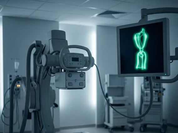
A Fluoroscope is a medical imaging device that utilizes X-rays to create a continuous, moving image of the inside of a patient’s body. Unlike traditional X-rays, which produce static images, a fluoroscope allows clinicians to observe internal organs, bones, and tissues in motion. This real-time visualization is achieved by passing a continuous X-ray beam through the patient, which then strikes a fluorescent screen or image intensifier, converting the X-ray energy into a visible light image. This dynamic imaging capability is often referred to as Fluoroscope medical imaging explained, highlighting its ability to show physiological processes as they happen.
The images produced by a fluoroscope are displayed on a monitor, enabling medical professionals to watch the movement of contrast agents through blood vessels or digestive tracts, observe joint movement, or guide surgical instruments. The technology has evolved significantly since its invention, with modern fluoroscopes offering digital imaging, dose reduction features, and enhanced image clarity, making it a cornerstone of contemporary diagnostic and interventional medicine.
How Fluoroscopy Works and Its Medical Applications
How Fluoroscopy Works
Fluoroscopy operates on the principle of continuous X-ray exposure. When a patient is positioned between an X-ray source and an image detector, the X-rays pass through the body. Different tissues absorb X-rays at varying rates; for instance, bones absorb more X-rays than soft tissues. The X-rays that pass through the body strike an image intensifier or a flat-panel detector, which converts the X-ray energy into a visible image. This image is then processed and displayed on a monitor in real-time, creating a live video feed of the body’s internal structures. Often, a contrast agent, such as barium or iodine, is introduced into the body (e.g., swallowed, injected, or administered via enema) to highlight specific organs, blood vessels, or pathways, making them more visible on the fluoroscopic images.
Medical Applications of Fluoroscopy
Fluoroscopy is a highly versatile tool with numerous applications across various medical specialties. Its ability to provide real-time imaging makes it invaluable for both diagnostic purposes and guiding interventional procedures. Here are some common uses:
- Gastrointestinal Studies: Used for barium swallows, upper GI series, and barium enemas to visualize the esophagus, stomach, small intestine, and colon, helping diagnose conditions like ulcers, tumors, or strictures.
- Cardiovascular Procedures: Essential for angiography to visualize blood vessels, cardiac catheterization to assess heart function, and stent placement to open blocked arteries.
- Orthopedic Procedures: Guides joint injections, fracture reductions, and the placement of screws or pins during orthopedic surgeries.
- Urological Procedures: Employed in voiding cystourethrograms (VCUGs) to assess bladder and urethra function, and to guide urinary stent placements.
- Pain Management: Used to precisely guide needles for nerve blocks or epidural injections in the spine.
- Foreign Body Retrieval: Helps in locating and guiding the removal of foreign objects from the body.
Benefits and Risks of Fluoroscopy
The use of fluoroscopy offers significant benefits in medical diagnostics and interventions, primarily due to its real-time imaging capability. It allows physicians to observe dynamic processes, such as the movement of the heart or the flow of contrast through vessels, which static imaging cannot provide. This immediate feedback is crucial for guiding complex procedures, ensuring precise placement of catheters, stents, or other devices, and reducing the need for repeat interventions. For example, in cardiology, fluoroscopy enables accurate navigation through delicate blood vessels, improving patient outcomes. The ability to visualize internal structures in motion also aids in more accurate diagnoses of functional abnormalities that might be missed with static imaging.
However, like all procedures involving ionizing radiation, fluoroscopy carries certain risks. The primary concern is radiation exposure, which can slightly increase the lifetime risk of cancer. The amount of radiation exposure varies depending on the duration of the procedure, the area of the body being examined, and the equipment used. Modern fluoroscopy systems are designed with dose-reduction features, and medical professionals are trained to use the lowest possible radiation dose (ALARA principle: As Low As Reasonably Achievable) while still obtaining diagnostic quality images. According to the American College of Radiology, the benefits of fluoroscopy generally outweigh the potential risks when medically indicated and performed by qualified personnel. Other potential risks, though rare, include skin irritation or burns from prolonged exposure, particularly in lengthy interventional procedures, and allergic reactions to contrast agents.