Fluorine F 18 Sodium Fluoride Pet
Fluorine F 18 Sodium Fluoride PET is an advanced imaging technique used in medical diagnostics, primarily for assessing bone metabolism and detecting skeletal abnormalities. This scan offers detailed insights into bone activity, aiding in the diagnosis and staging of various conditions.
Key Takeaways
- Fluorine F 18 Sodium Fluoride PET is a highly sensitive imaging scan primarily used for evaluating bone health.
- It utilizes a radioactive tracer, Fluorine-18 sodium fluoride, which accumulates in areas of increased bone turnover.
- The scan is particularly effective in detecting bone metastases, fractures, and other metabolic bone diseases.
- Preparation typically involves fasting and avoiding certain medications, with the procedure being non-invasive.
- Results provide crucial information for diagnosis, treatment planning, and monitoring disease progression.
What is Fluorine F 18 Sodium Fluoride PET (F-18 NaF PET)?
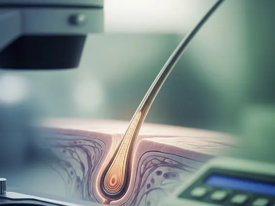
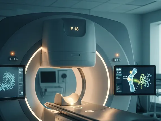
Fluorine F 18 Sodium Fluoride PET (F-18 NaF PET) is a sophisticated nuclear medicine imaging technique that provides highly detailed images of bone metabolism. It involves the intravenous injection of a small amount of a radioactive tracer, Fluorine-18 sodium fluoride (F-18 NaF). This tracer behaves similarly to calcium and is rapidly incorporated into areas of active bone formation and remodeling. The positron emission tomography (PET) scanner then detects the gamma rays emitted by the decaying F-18, creating precise images that highlight regions of increased bone turnover.
This imaging modality offers superior sensitivity and spatial resolution compared to traditional bone scans, making it invaluable for detecting subtle changes in bone activity. The detailed F-18 NaF PET imaging allows clinicians to visualize metabolic processes within the bone, providing an earlier and more accurate assessment of various skeletal conditions. The rapid clearance of the tracer from soft tissues enhances image contrast, further improving diagnostic accuracy.
What is Fluorine-18 Sodium Fluoride PET Scan Used For?
The Fluorine-18 sodium fluoride PET scan uses are diverse, primarily focusing on conditions affecting bone metabolism and structure. Its high sensitivity makes it particularly useful in oncology for detecting bone metastases, often before they are visible on conventional imaging. Beyond cancer, it plays a significant role in evaluating other bone-related disorders. The comprehensive Sodium fluoride F18 PET scan information it provides helps in accurate diagnosis and treatment planning.
Common applications for an F-18 NaF PET scan include:
- Detection of Bone Metastases: Identifying the spread of cancer to the bones, especially from primary tumors like prostate, breast, and lung cancer. It can detect lesions earlier than conventional bone scintigraphy.
- Evaluation of Primary Bone Tumors: Assessing the extent and metabolic activity of both benign and malignant bone tumors.
- Assessment of Metabolic Bone Diseases: Diagnosing and monitoring conditions such as Paget’s disease, osteomyelitis (bone infection), and complex regional pain syndrome.
- Identification of Fractures: Detecting occult fractures, stress fractures, or non-union of fractures that may not be clearly visible on X-rays or other imaging.
- Monitoring Treatment Response: Evaluating the effectiveness of therapies for bone metastases or other bone conditions by observing changes in bone metabolic activity.
The detailed F18 sodium fluoride PET scan explanation of bone activity helps clinicians make informed decisions about patient care, improving outcomes by guiding targeted treatments.
Preparing for and Undergoing an F-18 NaF PET Scan
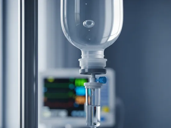
Preparing for an F-18 NaF PET scan is relatively straightforward, but adherence to specific instructions is crucial for optimal image quality. Patients are typically advised to fast for a few hours before the scan, usually 4-6 hours, to ensure accurate tracer distribution. Hydration is encouraged, but sugary drinks should be avoided. Patients should also inform their doctor about any medications they are taking, as some might need to be temporarily discontinued. It’s important to avoid strenuous physical activity for 24 hours prior to the scan, as this can alter tracer uptake in muscles.
On the day of the scan, patients will receive an intravenous injection of the F-18 NaF tracer. After the injection, there is typically a waiting period of 60-90 minutes to allow the tracer to circulate and accumulate in the bones. During this time, patients are encouraged to rest quietly and minimize movement. Following the uptake period, the patient will lie on a scanning bed that moves slowly through the PET scanner. The scan itself usually takes between 20 to 40 minutes, depending on the area being imaged. The procedure is generally well-tolerated and non-invasive, with minimal discomfort. After the scan, patients can typically resume their normal activities, with advice to drink plenty of fluids to help flush the remaining tracer from their system.