Fluorescence In Situ Hybridization
Fluorescence In Situ Hybridization is a powerful laboratory technique used to visualize and map genetic material in an individual’s cells, playing a critical role in the diagnosis and understanding of various genetic conditions and diseases.
Key Takeaways
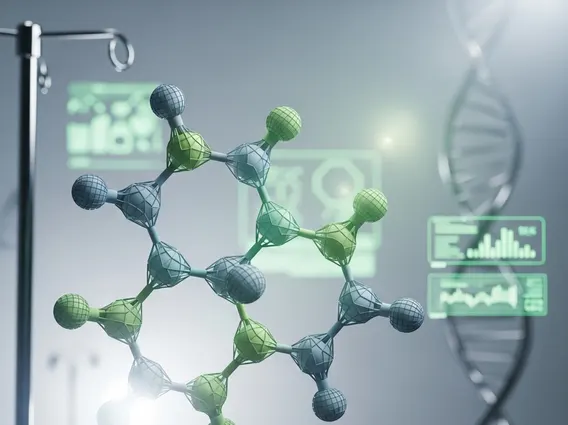
- Fluorescence In Situ Hybridization (FISH) is a molecular cytogenetic technique that uses fluorescent probes to detect specific DNA or RNA sequences within cells.
- The technique involves denaturing cellular DNA, allowing fluorescently labeled probes to bind to complementary target sequences.
- FISH is widely applied in medical diagnostics, including oncology for cancer staging and prognosis, and in prenatal diagnosis for detecting chromosomal abnormalities.
- It provides rapid and precise identification of genetic changes that are often too subtle to be detected by conventional microscopy.
What is Fluorescence In Situ Hybridization (FISH)?
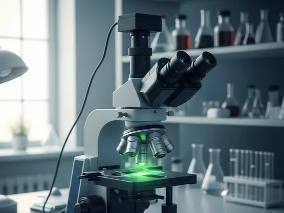
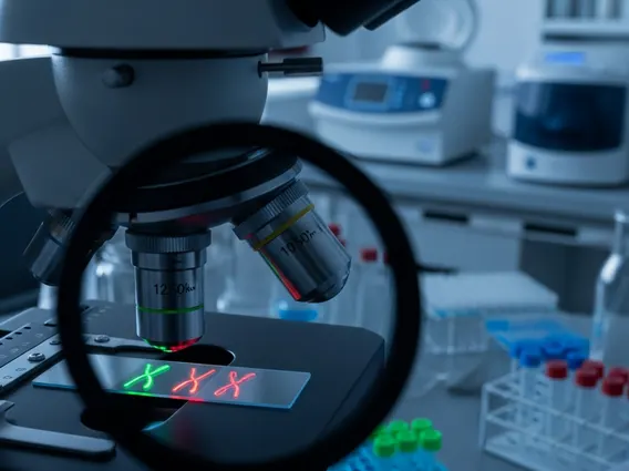
Fluorescence In Situ Hybridization (FISH) is a laboratory method used to detect and locate a specific DNA or RNA sequence on a chromosome. This molecular cytogenetic technique employs fluorescent probes that bind only to parts of the chromosome with a high degree of sequence complementarity. The fluorescent signals are then visualized under a fluorescence microscope, allowing researchers and clinicians to identify specific genetic alterations, such as gene amplifications, deletions, or translocations.
FISH has revolutionized the field of genetic diagnostics by providing a precise and sensitive way to analyze the genetic makeup of cells. Unlike traditional karyotyping, which relies on visible chromosomal banding patterns, FISH can pinpoint much smaller genetic changes and can be performed on various types of cells, including those from blood, bone marrow, tissue biopsies, and even amniotic fluid.
How Fluorescence In Situ Hybridization Works
The underlying principle of FISH relies on the ability of a single-stranded nucleic acid probe to hybridize (bind) to its complementary target sequence within the cellular DNA or RNA. The process involves several key steps, making it a robust FISH technique explanation for genetic analysis.
The general workflow for FISH includes:
- Sample Preparation: Cells are typically fixed onto a microscope slide, and their nuclei are permeabilized to allow probe entry.
- Probe Hybridization: Fluorescently labeled DNA or RNA probes, designed to be complementary to the target genetic sequence, are applied to the prepared cells. Both the cellular DNA and the probes are denatured (separated into single strands) by heat.
- Binding and Washing: As the sample cools, the probes hybridize (bind) to their specific target sequences on the chromosomes. Unbound probes are then washed away to minimize background noise.
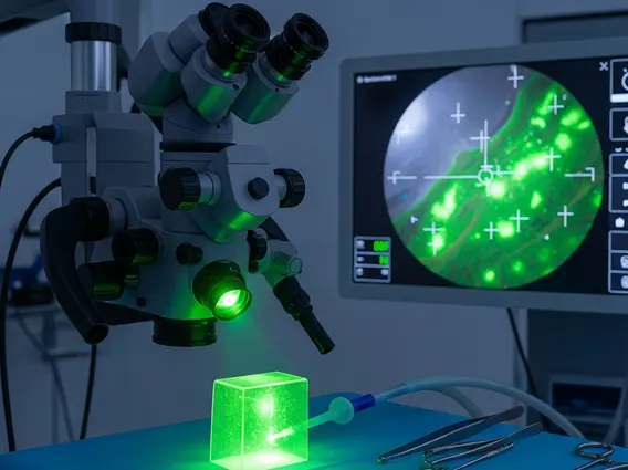
- Visualization: The slide is viewed under a fluorescence microscope, which excites the fluorescent dyes on the probes. The specific location and intensity of the fluorescent signals indicate the presence, absence, or rearrangement of the target genetic material.
This technique allows for the precise mapping of genes and the detection of chromosomal aberrations that are often too small to be seen with standard cytogenetic methods.
Applications of Fluorescence In Situ Hybridization
The Applications of Fluorescence In Situ Hybridization are extensive and continue to expand across various medical and research fields due to its high specificity and sensitivity. FISH has become an indispensable tool in clinical diagnostics, particularly in oncology and prenatal care.
In oncology, FISH is crucial for diagnosing and monitoring various cancers. For instance, it is widely used to detect the HER2 gene amplification in breast cancer, which guides treatment decisions for targeted therapies. Similarly, FISH can identify the Philadelphia chromosome (a translocation between chromosomes 9 and 22) in chronic myeloid leukemia (CML), which is a key diagnostic and prognostic marker. According to the American Cancer Society, accurate staging and molecular profiling, often aided by FISH, are vital for effective cancer management.
Beyond cancer, FISH plays a significant role in prenatal diagnosis, allowing for the rapid detection of common chromosomal aneuploidies such as trisomy 21 (Down syndrome), trisomy 18 (Edwards syndrome), and trisomy 13 (Patau syndrome) from amniotic fluid or chorionic villus samples. It is also used in postnatal diagnosis to identify microdeletion syndromes and other genetic disorders in children and adults, providing critical information for genetic counseling and patient management.