Fiducial Marker
A fiducial marker is a small, implantable object used in medical imaging and radiation therapy to precisely identify and track the position of a target area within the body. These markers serve as reference points, enhancing the accuracy of treatment delivery and diagnostic procedures.

Key Takeaways
- Fiducial markers are small, inert implants used to pinpoint specific anatomical locations during medical procedures.
- They significantly improve the precision of radiation therapy by allowing real-time tracking of tumor movement.
- Markers are typically made from radiopaque materials like gold, visible on imaging scans such as X-rays, CT, and MRI.
- Their primary function is to guide targeted treatments, minimizing damage to surrounding healthy tissue.
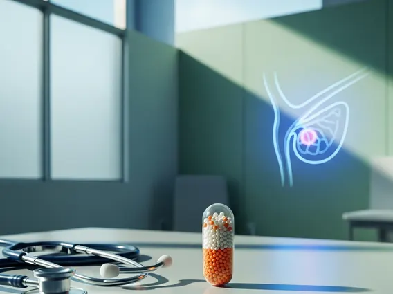
- Common clinical applications include prostate, lung, and liver cancer treatments, where organ motion can be a challenge.
What is a Fiducial Marker?
A fiducial marker refers to a small, inert object implanted into a patient’s body to provide a clear, visible reference point during medical imaging and treatment. These markers are crucial for enhancing the accuracy and precision of various medical procedures, particularly in oncology. The primary purpose of a fiducial marker is to delineate the exact location of a tumor or other target tissue, especially in areas prone to movement due to respiration, digestion, or other physiological processes.
The fiducial marker definition uses extend across diagnostic imaging, surgical guidance, and most notably, radiation therapy. By providing a consistent and easily identifiable point on imaging scans, these markers enable clinicians to precisely target treatment beams or surgical instruments, thereby minimizing damage to surrounding healthy tissues and organs. This precision is vital for maximizing treatment efficacy and reducing potential side effects for the patient.
Mechanism and Function of Fiducial Markers
Fiducial markers work by being highly visible on medical imaging scans, acting as beacons for the target area. Once implanted, typically through a minimally invasive procedure, these markers become integrated with the tissue. During imaging sessions, such as computed tomography (CT), magnetic resonance imaging (MRI), or X-ray fluoroscopy, the radiopaque nature of most markers allows them to stand out clearly against the surrounding soft tissues, which might otherwise be difficult to distinguish.
The mechanism by which fiducial markers function is critical for advanced radiation therapy techniques like image-guided radiation therapy (IGRT) and stereotactic body radiation therapy (SBRT). These techniques rely on real-time tracking of the target. The markers allow the radiation delivery system to adjust for any movement of the tumor, ensuring that the radiation dose is delivered precisely to the intended area. For example, in lung cancer treatment, a tumor can shift with every breath; fiducial markers enable the radiation beam to track this movement, maintaining accuracy. According to the American Society for Radiation Oncology (ASTRO), precision in radiation delivery is paramount for effective cancer treatment and reducing toxicity to healthy tissues, a goal significantly aided by fiducial markers.
Types and Clinical Uses of Fiducial Markers
There are several types of fiducial markers, primarily distinguished by their material composition, size, and shape, each chosen based on the specific clinical application and imaging modality. The most common materials are gold, carbon, and stainless steel, selected for their biocompatibility and high visibility on imaging scans. Gold markers, for instance, are widely used due to their excellent radiopacity and inertness within the body. They come in various forms, including tiny coils, spheres, or rods, designed to be easily implanted and remain stable.
The clinical uses of fiducial markers are diverse and continue to expand, particularly in the field of oncology. They are indispensable for treating cancers in organs that are subject to movement or are located near critical structures. Key applications include:
- Prostate Cancer: Prostate glands can shift significantly, making markers essential for accurate radiation targeting and reducing rectal toxicity.
- Lung Cancer: Tumors in the lungs move with respiration, and markers enable real-time tracking and gating of radiation delivery.
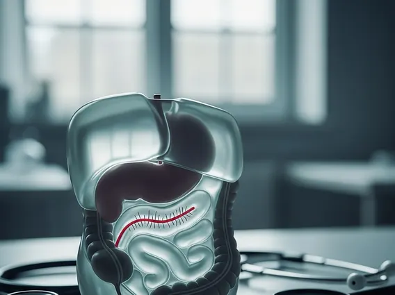
- Liver Cancer: Similar to lung tumors, liver lesions can move, requiring markers for precise SBRT.
- Pancreatic Cancer: Markers help delineate the tumor from surrounding sensitive organs like the duodenum.
- Breast Cancer: Used to mark the tumor bed after lumpectomy to guide subsequent radiation therapy.
These markers facilitate highly conformal radiation delivery, allowing clinicians to administer higher, more effective doses to the tumor while sparing adjacent healthy tissues, ultimately improving patient outcomes and quality of life.