
Extremely Dense Breast Tissue
Extremely dense breast tissue is a common finding on mammograms, characterized by a higher proportion of glandular and fibrous tissue compared to fatty tissue. This condition has significant implications for breast cancer screening and risk assessment, necessitating a tailored approach to women’s health.
Key Takeaways
- Extremely Dense Breast Tissue refers to breasts with a high amount of glandular and fibrous tissue, making them appear white on mammograms.
- This density can obscure tumors on standard mammograms, making early detection more challenging.
- Women with extremely dense breast tissue have an elevated risk of developing breast cancer compared to those with less dense breasts.
- There are typically no discernible symptoms of extremely dense breast tissue; it is identified through mammography.
- Enhanced screening strategies, such as supplemental imaging, are often recommended for managing extremely dense breast tissue.
What is Extremely Dense Breast Tissue?
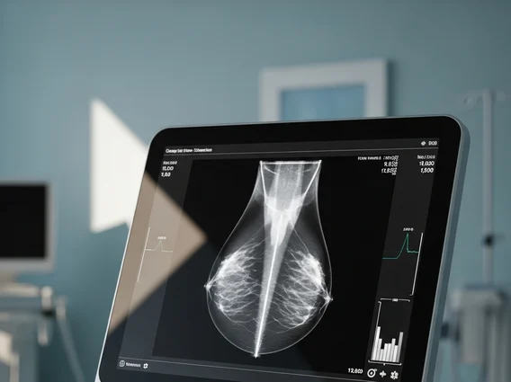
Extremely Dense Breast Tissue refers to breasts composed predominantly of glandular and fibrous connective tissue with very little fat. This is the highest category of breast density, often classified as BI-RADS D on a mammogram. Unlike fatty tissue, which appears dark and transparent on a mammogram, dense tissue appears white, similar to how tumors appear. This characteristic makes it challenging to detect potential cancers, as abnormalities can be masked by the surrounding dense tissue.
Breast density is a common trait, affecting approximately 40-50% of women in the United States, with about 10% having extremely dense breasts. The exact cause of varying breast density is not fully understood, but factors such as genetics, age (density tends to decrease with age, though not always), hormonal status, and certain medications can play a role. It is important to note that breast density is not determined by how breasts feel during a physical exam; it can only be assessed through a mammogram.
Extremely Dense Breast Tissue Risks and Implications
Having extremely dense breast tissue risks are primarily twofold: it can obscure cancers on mammograms and it is an independent risk factor for developing breast cancer. The masking effect occurs because both dense tissue and cancerous tumors appear white on a mammogram, making it difficult for radiologists to distinguish between them. This can lead to delayed detection of breast cancer, potentially allowing tumors to grow larger before they are found.
Furthermore, studies indicate that women with extremely dense breasts have a significantly higher risk of developing breast cancer compared to women with fatty breasts. For instance, women with extremely dense breasts may have a four to six times higher risk of breast cancer compared to those with fatty breasts, according to the American Cancer Society. It is crucial to understand that there are generally no specific symptoms of extremely dense breast tissue that a woman can feel; the condition is asymptomatic and identified solely through mammographic imaging. Therefore, women are often unaware of their breast density until informed by their healthcare provider after a mammogram.
Managing Extremely Dense Breast Tissue: Screening Guidelines
For women with extremely dense breast tissue, standard mammography alone may not be sufficient for effective breast cancer screening. Therefore, healthcare providers often recommend enhanced screening strategies for managing extremely dense breast tissue. These supplemental screenings aim to overcome the limitations of mammography in dense breasts and improve the chances of early cancer detection.
Common supplemental screening methods include:
- Breast Ultrasound: This imaging technique uses sound waves to create images of breast tissue. It is particularly effective at detecting small cancers in dense breasts that might be missed by mammography, as it does not rely on tissue density for image clarity.
- Magnetic Resonance Imaging (MRI): Breast MRI uses magnetic fields and radio waves to create detailed images. It is highly sensitive in detecting breast cancers in dense breasts but is typically reserved for women with additional high-risk factors due to its higher cost and potential for false positives.
- 3D Mammography (Tomosynthesis): This advanced form of mammography takes multiple images from different angles, creating a 3D reconstruction of the breast. It can help radiologists see through dense tissue layers more effectively, reducing the masking effect and improving cancer detection rates compared to conventional 2D mammography.
It is essential for women with extremely dense breast tissue to discuss their personal risk factors and screening options with their healthcare provider to develop a personalized screening plan. This proactive approach, combining regular mammograms with appropriate supplemental imaging, is key to optimizing early detection and improving outcomes.


