
Electrocardiogram
An Electrocardiogram is a common and vital diagnostic tool used in medicine to assess the electrical activity of the heart. This non-invasive test provides crucial insights into cardiac function, helping healthcare professionals identify various heart conditions.
Key Takeaways
- An Electrocardiogram (ECG or EKG) records the heart’s electrical signals.
- It is a quick, painless, and non-invasive procedure.
- The test helps diagnose a range of heart conditions, including arrhythmias and heart attacks.
- Electrodes placed on the skin detect electrical impulses from the heart.
- Interpretation involves analyzing specific waves and intervals on the tracing.
What is an Electrocardiogram (ECG) and Its Purpose?
An Electrocardiogram (ECG or EKG) is a medical test that measures and records the electrical activity of the heart over a period of time. It translates the heart’s electrical impulses into a wave pattern displayed on paper or a monitor. This diagnostic tool is fundamental in cardiology, offering a snapshot of how the heart is functioning electrically.
The primary purpose of an ECG test is to detect and evaluate various heart conditions. It can identify irregular heart rhythms (arrhythmias), determine if a heart attack has occurred or is in progress, assess the effectiveness of certain heart medications, and check for issues like an enlarged heart or inflammation of the heart sac (pericarditis). The simplicity and speed of an ECG make it an indispensable first-line diagnostic test in both emergency and routine clinical settings.
How Does an Electrocardiogram (EKG) Work?
An EKG works by detecting the tiny electrical impulses generated by the heart as it beats. During the procedure, small adhesive electrodes are placed on specific areas of the chest, arms, and legs. These electrodes are connected by wires to an EKG machine, which records the electrical activity. The heart’s electrical system initiates each heartbeat, starting with a signal from the sinoatrial (SA) node, which then spreads through the atria and ventricles, causing them to contract and pump blood.
The EKG machine captures these electrical signals from different angles, providing a comprehensive view of the heart’s electrical pathway. It amplifies these signals and converts them into a visual tracing. This tracing shows a series of waves and deflections that correspond to the different phases of the heart’s electrical cycle, such as atrial contraction, ventricular contraction, and ventricular relaxation. The entire process is quick, typically taking only a few minutes, and involves no discomfort beyond the placement of the electrodes.
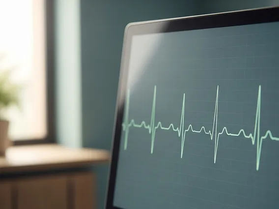
Electrocardiogram Interpretation Basics
Understanding electrocardiogram interpretation basics involves recognizing the characteristic waves, intervals, and segments on the EKG tracing, and assessing their timing, amplitude, and morphology. A standard EKG tracing displays a repeating pattern that reflects the heart’s electrical cycle. Deviations from this normal pattern can indicate underlying cardiac issues.
Key components analyzed during interpretation include:
- P wave: Represents atrial depolarization, which is the electrical activation of the atria, leading to their contraction.
- QRS complex: Represents ventricular depolarization, the electrical activation of the ventricles, leading to their contraction and blood ejection.
- T wave: Represents ventricular repolarization, the electrical recovery of the ventricles, allowing them to relax and refill with blood.
- PR interval: Measures the time from the beginning of atrial activation to the beginning of ventricular activation, indicating the conduction time through the AV node.
- ST segment: The segment between the QRS complex and the T wave, which is critical for identifying myocardial ischemia or injury.
By carefully examining these components and their relationships, healthcare professionals can diagnose a wide range of cardiac conditions, from minor rhythm disturbances to life-threatening heart attacks. The precise measurement of intervals and segments, along with the shape and direction of the waves, provides vital diagnostic information.


