
Ekg
Ekg, more commonly known as an electrocardiogram, is a fundamental diagnostic tool used to assess the electrical activity of the heart. This non-invasive test provides crucial insights into cardiac health, helping medical professionals detect and monitor various heart conditions.
Key Takeaways
- An Electrocardiogram (EKG) records the heart’s electrical signals to detect cardiac abnormalities.
- The EKG machine uses electrodes placed on the skin to capture these signals, which are then displayed as a waveform.
- EKG readings help diagnose conditions like arrhythmias, heart attacks, and structural heart disease.
- The test is quick, painless, and a vital component of cardiology diagnostics.
- Interpreting EKG results requires specialized medical knowledge to identify specific patterns.
What is an EKG (Electrocardiogram) and What Does It Show?
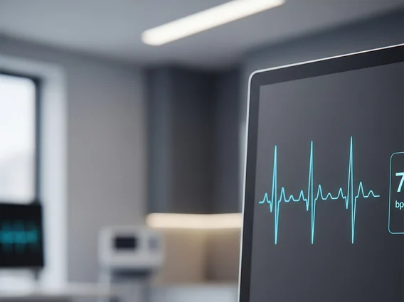
Electrocardiogram (EKG) is a non-invasive medical test that records the electrical signals of the heart. These electrical impulses coordinate the contraction and relaxation of the heart chambers, pumping blood throughout the body. By measuring these signals, an EKG provides a detailed graphical representation of the heart’s rhythm and electrical activity. The purpose of EKG test in cardiology is primarily to evaluate heart function, detect abnormalities, and guide treatment decisions for a wide range of cardiac conditions.
An EKG can reveal several important aspects of heart health. It shows how fast the heart is beating and whether its rhythm is steady or irregular (arrhythmia). It can also indicate if parts of the heart are enlarged, overworked, or damaged due to conditions like a heart attack or ischemia. Furthermore, an EKG can identify issues with the heart’s conduction system, which is responsible for transmitting electrical signals efficiently. This comprehensive view makes it an indispensable tool for diagnosing and monitoring cardiovascular diseases.
How Does an EKG Machine Work?
An EKG machine works by detecting and amplifying the tiny electrical currents generated by the heart, then translating them into a visual waveform. The process begins with the placement of small, sticky electrodes on specific locations on the patient’s chest, arms, and legs. These electrodes are connected by wires to the EKG machine. Each electrode acts as a sensor, picking up the electrical impulses as they travel through the heart muscle.
Once the electrodes are in place, the EKG machine records these electrical signals over a short period, typically a few minutes. The machine processes the signals and prints them out on a grid paper or displays them on a screen. The resulting tracing, known as an electrocardiogram, consists of a series of waves and segments that correspond to different phases of the heart’s electrical cycle. For instance, the P wave represents atrial depolarization, the QRS complex represents ventricular depolarization, and the T wave represents ventricular repolarization. This precise recording allows cardiologists to analyze the timing and strength of each electrical event.
- Electrode Placement: Up to 12 electrodes are strategically placed on the skin.
- Signal Detection: Electrodes detect the heart’s electrical activity.
- Amplification & Recording: The EKG machine amplifies and records these signals.
- Waveform Generation: Signals are converted into a visual tracing on paper or screen.
Understanding EKG Results and Readings
Interpreting EKG results and readings requires specialized training and expertise. A cardiologist or other trained medical professional analyzes the various components of the EKG waveform to identify any deviations from normal patterns. The key elements examined include the heart rate, rhythm, and the shape, duration, and amplitude of the P wave, QRS complex, and T wave, as well as the intervals between them.
For example, an abnormally fast heart rate (tachycardia) or slow heart rate (bradycardia) can be easily identified. Irregular rhythms, known as arrhythmias, can manifest in various forms, such as atrial fibrillation or ventricular tachycardia, each with distinct EKG signatures. Changes in the ST segment or T wave can indicate myocardial ischemia (reduced blood flow to the heart muscle) or a heart attack. According to the Centers for Disease Control and Prevention (CDC), heart disease remains a leading cause of death, making early and accurate diagnosis through tools like EKG critical for timely intervention.
| EKG Wave/Segment | Represents | Potential Abnormalities |
|---|---|---|
| P Wave | Atrial depolarization (contraction) | Enlarged atria, abnormal atrial rhythms |
| QRS Complex | Ventricular depolarization (contraction) | Ventricular hypertrophy, bundle branch blocks, heart attack |
| T Wave | Ventricular repolarization (relaxation) | Ischemia, electrolyte imbalances, certain medications |
| PR Interval | Time from atrial to ventricular contraction | Heart blocks |
| ST Segment | Time between ventricular contraction/relaxation | Myocardial ischemia, heart attack, pericarditis |
This table provides a simplified overview; a comprehensive interpretation considers all these elements in context with the patient’s clinical picture. Regular EKG screenings, especially for individuals with risk factors for heart disease, can play a significant role in preventive care and early detection.


