Echocardiography
Echocardiography is a vital diagnostic tool in cardiology, providing detailed images of the heart’s structure and function. This non-invasive procedure helps clinicians assess various cardiac conditions, guiding treatment decisions and improving patient outcomes.
Key Takeaways
- Echocardiography uses sound waves to create real-time images of the heart.
- It assesses heart chambers, valves, blood flow, and overall cardiac function.
- Different types, such as transthoracic and transesophageal, offer varying levels of detail.
- The procedure is non-invasive, generally painless, and crucial for diagnosing many heart conditions.
- Preparation is minimal, and results help guide personalized treatment plans.
What is Echocardiography?
Echocardiography refers to a diagnostic medical test that uses high-frequency sound waves (ultrasound) to produce live images of the heart. These images, called echocardiograms, allow doctors to visualize the heart’s chambers, valves, major blood vessels, and the sac surrounding the heart (pericardium). It provides crucial information about the heart’s size, shape, and pumping ability, as well as the presence of any damage or abnormalities. This non-invasive procedure is fundamental in diagnosing and monitoring a wide range of cardiac conditions.
According to the American Heart Association, heart disease remains a leading cause of death globally, underscoring the importance of diagnostic tools like echocardiography for early detection and management.
How Echocardiography Works and Its Different Types
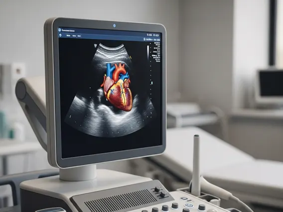
How echocardiography works involves a transducer, a small handheld device, which emits sound waves that travel through the chest to the heart. These sound waves bounce off the heart structures and return to the transducer, which then sends them to a computer. The computer processes these echoes into real-time moving images displayed on a monitor. This allows cardiologists to observe the heart beating, blood flowing through its chambers and valves, and assess its overall function dynamically.
There are several types of echocardiography, each suited for different diagnostic needs:
- Transthoracic Echocardiography (TTE): This is the most common type. The transducer is placed on the chest wall, and images are obtained by moving it across the skin. It’s non-invasive and provides a general overview of heart structure and function.
- Transesophageal Echocardiography (TEE): For more detailed images, especially of the back of the heart or when TTE is unclear, a TEE may be performed. A smaller transducer is attached to a thin tube that is guided down the esophagus (the tube connecting the mouth to the stomach). Because the esophagus is close to the heart, TEE provides clearer images without interference from the ribs or lungs.
- Stress Echocardiography: This type is performed before and immediately after physical exercise (on a treadmill or stationary bike) or after administering medication that simulates the effects of exercise on the heart. It helps evaluate how the heart functions under stress and can detect reduced blood flow to the heart muscle.
- Doppler Echocardiography: Often used in conjunction with TTE or TEE, Doppler technology measures the speed and direction of blood flow through the heart and blood vessels. This helps detect abnormal blood flow patterns, such as those caused by narrowed or leaky heart valves.
- 3D Echocardiography: This advanced technique provides three-dimensional views of the heart, offering more detailed anatomical information, particularly useful for complex congenital heart defects or planning surgical interventions.
The Echocardiography Procedure Explained
The echocardiography procedure explained typically involves minimal preparation, especially for a standard transthoracic echocardiogram. Patients are usually asked to wear loose clothing and may be asked to remove jewelry from the neck or chest area. For TEE, patients will need to fast for several hours before the procedure.
During a standard transthoracic echocardiogram:
- The patient lies on an examination table, often on their left side.
- A sonographer applies a special gel to the chest, which helps the transducer make good contact with the skin and facilitates the transmission of sound waves.
- The sonographer moves the transducer across different areas of the chest to capture images from various angles.
- Patients may be asked to hold their breath briefly or change positions to improve image quality.
- The procedure is generally painless and takes about 30 to 60 minutes.
After the procedure, the gel is wiped off, and patients can typically resume their normal activities immediately. The images are then reviewed by a cardiologist, who will interpret the findings and discuss them with the patient or their referring physician. The results of an echocardiogram are crucial for diagnosing conditions such as heart valve disease, heart failure, congenital heart defects, and issues with the heart muscle, guiding appropriate treatment strategies.