Diagnosis, Screening, and Early Detection of Melanoma
Melanoma, a serious form of skin cancer, can be life-threatening if not identified and treated promptly. Understanding the processes of melanoma diagnosis, effective screening methods, and the importance of early detection is crucial for improving patient outcomes.

Key Takeaways
- Early detection of melanoma significantly improves treatment success and survival rates.
- Regular self-skin exams using the ABCDE rule are vital for recognizing potential signs of early melanoma.
- Professional skin screenings, especially for individuals at higher risk, are a key component of melanoma screening guidelines.
- Melanoma diagnosis involves visual examination, dermoscopy, and ultimately, a biopsy for pathological confirmation.
- Prompt consultation with a dermatologist for any suspicious skin changes is paramount.
What is Melanoma Diagnosis?
What is melanoma diagnosis refers to the comprehensive process of identifying the presence of melanoma, determining its type, and assessing its stage. This process begins with a thorough evaluation of a suspicious lesion and culminates in a definitive pathological confirmation, which is essential for guiding subsequent treatment decisions.
Initial Assessment and Patient History
The initial step in melanoma diagnosis involves a detailed patient history and a physical examination. Healthcare providers will inquire about personal and family history of melanoma, excessive sun exposure, tanning bed use, and the presence of numerous or atypical moles. They will also ask about any changes observed in existing moles or the appearance of new, unusual skin lesions. This information helps assess an individual’s risk factors and guides the subsequent examination of the skin.
Purpose of a Definitive Diagnosis
The primary purpose of obtaining a definitive melanoma diagnosis is to confirm the presence of cancerous cells and to differentiate melanoma from benign lesions. An accurate diagnosis is fundamental for determining the appropriate treatment strategy, which can range from surgical excision for early-stage melanoma to more complex therapies for advanced cases. Without a precise diagnosis, effective treatment cannot be initiated, underscoring the critical role of pathology in confirming the disease.
How Melanoma is Diagnosed: Key Techniques
Understanding how is melanoma diagnosed involves a combination of visual assessment and advanced diagnostic procedures. These melanoma detection techniques are designed to identify suspicious lesions and confirm the presence of cancerous cells, ensuring an accurate and timely diagnosis.
Visual Examination and Dermoscopy
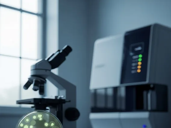
The diagnostic process often begins with a visual examination of the skin by a dermatologist. They meticulously inspect the entire skin surface, including areas not typically exposed to the sun, for any suspicious moles, growths, or lesions. A key tool in this stage is dermoscopy, a non-invasive technique that uses a handheld microscope to examine skin lesions with magnification and specialized lighting. Dermoscopy allows the dermatologist to visualize structures and patterns beneath the skin’s surface that are not visible to the naked eye, significantly improving the accuracy of identifying potential melanomas and other skin cancers without immediate biopsy.
Biopsy Procedures and Pathology
If a lesion appears suspicious after visual and dermoscopic examination, a biopsy is performed to obtain tissue for pathological analysis. This is the definitive step in how is melanoma diagnosed. There are several types of biopsies:
- Excisional Biopsy: The entire suspicious lesion and a small margin of surrounding healthy skin are removed. This is often preferred for suspected melanoma as it allows for complete assessment of the lesion’s depth.
- Incisional Biopsy: Only a part of the lesion is removed, typically when the lesion is very large or located in a cosmetically sensitive area.
- Punch Biopsy: A small, circular piece of the lesion is removed using a specialized tool.
The tissue sample is then sent to a pathologist, who examines it under a microscope to confirm the presence of melanoma cells, determine the type of melanoma, and assess its Breslow depth (thickness), which is a critical prognostic factor.
Recognizing Signs of Early Melanoma
Identifying signs of early melanoma is paramount for successful treatment. Both self-examination and professional checks play vital roles in melanoma early detection methods, empowering individuals to seek timely medical attention for suspicious changes.
The ABCDE Rule for Self-Checks
The ABCDE rule is a widely recognized and effective tool for self-monitoring moles and detecting potential signs of early melanoma. Regularly checking your skin for these characteristics can help identify suspicious lesions:
- A – Asymmetry: One half of the mole does not match the other half.
- B – Border Irregularity: The edges are ragged, notched, blurred, or poorly defined.
- C – Color Variation: The mole has uneven color, with shades of brown, black, tan, red, white, or blue.
- D – Diameter: The mole is typically larger than 6 millimeters (about the size of a pencil eraser), though melanomas can sometimes be smaller.
- E – Evolving: The mole is changing in size, shape, color, elevation, or any new symptoms like bleeding, itching, or crusting.
Any mole exhibiting one or more of these characteristics warrants immediate professional evaluation.
When to See a Dermatologist
It is crucial to consult a dermatologist if you notice any new moles, growths, or spots, or if existing ones begin to change in size, shape, color, or texture. Persistent itching, tenderness, bleeding, or non-healing sores on the skin should also prompt a visit to a specialist. Early intervention based on these melanoma early detection methods can significantly improve prognosis, as melanoma is highly treatable when caught in its initial stages.
Melanoma Screening Guidelines
Adhering to comprehensive melanoma screening guidelines is a proactive approach to safeguarding skin health, especially for individuals at increased risk. These guidelines encompass both self-monitoring and professional examinations to maximize the chances of early detection.
Who Needs Regular Skin Checks?
Regular skin checks are recommended for everyone, but certain individuals should follow more stringent melanoma screening guidelines due to elevated risk factors. These include people with a personal or family history of melanoma, those with numerous moles (especially atypical ones), individuals with fair skin that burns easily, a history of severe sunburns, or extensive exposure to UV radiation (from sun or tanning beds). Immunosuppressed individuals also face a higher risk. For these groups, annual or even more frequent professional skin examinations may be advised to ensure thorough surveillance.
Professional vs. Self-Screening
Both professional skin examinations by a dermatologist and regular self-screening are integral components of effective melanoma early detection methods. Self-screening allows individuals to monitor their skin for changes between professional visits, fostering a sense of personal responsibility for health. However, professional screenings offer the expertise of a dermatologist who can identify subtle or atypical lesions that might be missed during a self-exam, especially in hard-to-see areas like the scalp or back. Dermatologists use specialized tools like dermoscopes to examine suspicious lesions more closely. Combining both approaches provides the most comprehensive strategy for identifying melanoma at its earliest, most treatable stage.
Importance of Early Melanoma Detection
The importance of early melanoma diagnosis cannot be overstated, as it directly correlates with treatment efficacy and patient survival. Detecting melanoma when it is still confined to the top layers of the skin dramatically improves the chances of a complete cure.
Impact on Treatment and Prognosis
When melanoma is detected early, typically at Stage 0 or Stage I, it is often treated effectively with surgical excision alone. This involves removing the cancerous lesion and a small margin of healthy tissue, which is usually a straightforward procedure with minimal recovery time. In contrast, if melanoma progresses to later stages (Stage III or IV), it may have spread to lymph nodes or distant organs, requiring more aggressive and complex treatments such as immunotherapy, targeted therapy, chemotherapy, or radiation. These advanced treatments are often associated with more significant side effects and a lower probability of cure, highlighting the profound importance of early melanoma diagnosis in dictating the course and success of treatment.
Improving Survival Rates
The impact of early detection on survival rates is substantial. According to the National Cancer Institute’s SEER Program, the 5-year relative survival rate for localized melanoma (meaning it has not spread beyond the original site) is approximately 99%. However, this rate drops significantly to 74% if the melanoma has spread to regional lymph nodes and further to 35% if it has metastasized to distant parts of the body. These statistics powerfully illustrate why melanoma early detection methods are critical; they offer the best chance for long-term survival and underscore the urgency of regular screenings and prompt medical evaluation for any suspicious skin changes.
Frequently Asked Questions About Melanoma Diagnosis and Detection
How often should I check my skin for melanoma?
It is recommended to perform a thorough self-skin examination once a month, ideally after a shower or bath. Use a full-length mirror and a hand mirror to check all areas of your body, including your scalp, palms, soles, and between your toes. If you have a higher risk of melanoma, such as a family history or numerous moles, your dermatologist may recommend more frequent professional check-ups in addition to your self-exams.
Can melanoma be misdiagnosed?
While dermatologists are highly skilled, misdiagnosis can occasionally occur, either by mistaking a benign lesion for melanoma or vice versa. This is why a biopsy and pathological examination by a specialized dermatopathologist are crucial for a definitive melanoma diagnosis. The combination of visual assessment, dermoscopy, and expert pathology significantly reduces the risk of misdiagnosis, ensuring accurate identification and appropriate treatment.
What are the first steps if I find a suspicious mole?
If you discover a mole or skin lesion that exhibits any of the ABCDE characteristics, or if it is new, changing, itching, or bleeding, the immediate next step is to schedule an appointment with a dermatologist. Do not delay seeking professional medical advice. A dermatologist will perform a thorough examination and, if necessary, conduct a biopsy to determine if the lesion is cancerous, initiating prompt treatment if melanoma is confirmed.