Diagnosis, Screening, and Early Detection of Eye Cancer
Understanding the intricacies of eye cancer, from its subtle beginnings to comprehensive diagnostic approaches, is paramount for effective management. This article provides a detailed overview of how eye cancer is identified, emphasizing the critical role of early detection and timely intervention.
Key Takeaways
- Early Symptom Recognition: Be vigilant for visual changes like blurred vision, new floaters, or dark spots, as these can be signs of eye cancer early detection.
- Regular Eye Exams: Adhering to eye cancer screening guidelines through routine comprehensive eye examinations is crucial, especially for high-risk individuals.
- Advanced Diagnostic Methods: A definitive eye cancer diagnosis involves specialized ocular examinations, advanced imaging techniques, and sometimes a biopsy.
- Importance of Prompt Action: Swift identification and treatment significantly improve outcomes, preserving vision and overall health.
Recognizing Early Symptoms of Eye Cancer
Being aware of potential indicators is the first step in the early detection of eye cancer symptoms. While many symptoms can be attributed to less serious conditions, persistent or worsening signs warrant immediate medical attention. Understanding what are the symptoms of eye cancer can empower individuals to seek help promptly.
Visual Changes to Watch For
Many early indicators of eye cancer manifest as alterations in vision. These changes often affect one eye and can be subtle initially, making them easy to overlook. Recognizing these signs of eye cancer early detection is vital.
- Blurred Vision or Vision Loss: A gradual or sudden decrease in visual acuity in one eye, not corrected by glasses, can be a warning sign.
- New Floaters or Flashes of Light: An increase in the number of “floaters” (small specks or threads that drift across the field of vision) or experiencing flashes of light, especially if new or persistent, should be evaluated.
- Dark Spot on the Iris or Change in Pupil Shape: The appearance of a new, growing dark spot on the iris (the colored part of the eye) or an irregular pupil shape may indicate an underlying issue.
- Peripheral Vision Loss: A noticeable reduction in side vision can be a symptom, particularly if a tumor is growing in certain areas of the eye.
Physical Signs and Discomfort
Beyond visual disturbances, certain physical changes or sensations in and around the eye can also be indicative of eye cancer. While less common, these physical manifestations should not be ignored.
- Bulging of One Eye (Proptosis): If one eye appears to protrude more than the other, it could be a sign of a tumor growing behind the eyeball.
- Change in Eye Movement: Difficulty moving the eye in certain directions or a noticeable change in how the eyes track together can occur if a tumor affects the muscles surrounding the eye.
- Pain or Discomfort: Although eye cancer is often painless in its early stages, some types or advanced cases can cause localized pain, pressure, or a feeling of fullness in the eye.
- Redness or Irritation: Persistent redness, swelling, or irritation of the eye that doesn’t resolve with standard treatments may sometimes be a symptom.
Eye Cancer Screening Guidelines
Adherence to recommended eye cancer screening guidelines plays a pivotal role in identifying potential issues before they become advanced. Regular examinations allow eye care professionals to perform screening for eye cancer, even in the absence of noticeable symptoms.
Routine Eye Exams
Comprehensive eye exams are not just for checking vision; they are crucial for overall eye health, including the detection of various diseases. For the general population, routine eye exams are the primary method for screening for eye cancer.
- Frequency: Adults should generally have a comprehensive eye exam every one to two years, or as recommended by their eye care professional, especially after age 40. Children should also have regular screenings as advised by pediatricians.
- What They Involve: A thorough eye exam typically includes a visual acuity test, pupil dilation to allow examination of the retina and optic nerve, tonometry (to check eye pressure), and slit-lamp examination to inspect the front and back of the eye. An ophthalmologist or optometrist can identify suspicious lesions or abnormalities during these examinations.
- Early Detection: Many ocular cancers, such as melanoma, can be asymptomatic in their early stages. Routine exams provide an opportunity to detect these lesions when they are small and more treatable.
Targeted Screening for High-Risk Groups
Certain individuals face a higher risk of developing eye cancer due to genetic predispositions, specific medical conditions, or environmental factors. For these groups, more frequent or specialized screening for eye cancer is recommended.
High-risk factors that may necessitate targeted screening include:
- Family History: A strong family history of certain eye cancers, such as retinoblastoma or ocular melanoma, warrants closer monitoring.
- Genetic Syndromes: Individuals with specific genetic conditions like BAP1 tumor predisposition syndrome or neurofibromatosis type 1 have an increased risk.
- Excessive UV Exposure: Prolonged and unprotected exposure to ultraviolet (UV) radiation is a known risk factor for some types of eye cancer, including conjunctival melanoma.
- Certain Moles: People with numerous atypical moles on their skin, or a history of cutaneous melanoma, may have a higher risk of developing ocular melanoma.
- Prior Radiation Therapy: History of radiation to the head and neck can increase the risk of secondary cancers in the eye.
For these groups, an ophthalmologist may recommend more frequent check-ups, specialized imaging, or consultations with an ocular oncologist to ensure timely eye cancer diagnosis.
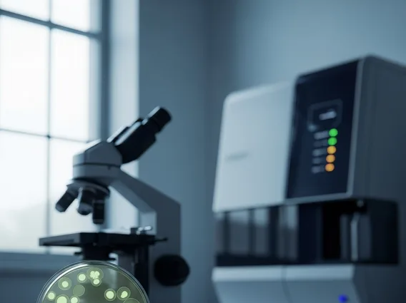
Comprehensive Eye Cancer Diagnosis Methods
When symptoms suggest a potential issue or a screening reveals an abnormality, a series of comprehensive eye cancer diagnosis methods are employed to confirm the presence of cancer, determine its type, and assess its extent. This multi-faceted approach helps answer the crucial question: how is eye cancer diagnosed definitively?
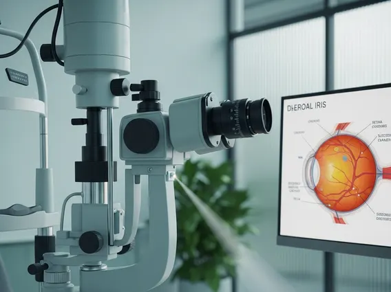
Ocular Examinations and Imaging
The diagnostic process typically begins with a detailed examination by an ophthalmologist, often one specializing in ocular oncology. This is followed by advanced imaging techniques that provide crucial insights without being invasive.
- Detailed Ophthalmoscopy: After dilating the pupils, the ophthalmologist uses specialized instruments (like an indirect ophthalmoscope or slit lamp with a high-power lens) to meticulously examine the inside of the eye, including the retina, choroid, and optic nerve. They look for tumors, abnormal blood vessels, or other suspicious lesions.
- Ocular Ultrasound (B-scan): This non-invasive test uses high-frequency sound waves to create detailed images of the eye’s internal structures, particularly useful for tumors located at the back of the eye or when the view is obscured by cataracts or hemorrhage. It can help determine the size, shape, and internal characteristics of a mass.
- Optical Coherence Tomography (OCT): OCT provides high-resolution cross-sectional images of the retina and choroid, helping to differentiate between benign lesions and malignant tumors, and to assess tumor thickness and retinal involvement.
- Fluorescein Angiography: A dye is injected into a vein, and a special camera takes pictures as the dye circulates through the blood vessels in the eye. This helps visualize abnormal blood vessel patterns associated with tumors.
- Magnetic Resonance Imaging (MRI) and Computed Tomography (CT) Scans: These advanced imaging techniques are used to assess the extent of the tumor, determine if it has spread outside the eyeball, or if it involves surrounding structures like the optic nerve or brain. MRI is particularly effective for soft tissues.
Biopsy and Pathology
While imaging can strongly suggest cancer, a biopsy is often the only way to confirm a definitive eye cancer diagnosis and determine the specific type of cancer. This involves taking a small tissue sample for microscopic examination by a pathologist.
- Fine-Needle Aspiration Biopsy (FNAB): For intraocular tumors (inside the eye), a very thin needle is used to extract a small sample of cells from the suspicious lesion. This is typically performed under local anesthesia. FNAB is often used for uveal melanoma to confirm the diagnosis and for genetic analysis, which can help predict prognosis.
- Incisional or Excisional Biopsy: For tumors on the surface of the eye (e.g., conjunctival melanoma) or on the eyelids, a small piece of the tumor (incisional) or the entire tumor (excisional) may be surgically removed for pathological analysis.
- Pathological Analysis: The tissue sample is examined under a microscope by a specialized pathologist. They identify cancerous cells, determine the type of cancer (e.g., melanoma, lymphoma, squamous cell carcinoma), and assess its characteristics, which are crucial for guiding treatment decisions.
The combination of these methods provides a comprehensive picture, allowing for an accurate eye cancer diagnosis and the formulation of an appropriate treatment plan.
The Importance of Early Detection and Treatment
The significance of early detection of eye cancer cannot be overstated. When eye cancer is identified in its nascent stages, the prognosis is generally far more favorable, leading to better treatment outcomes and a higher chance of preserving vision and the eye itself. Delay in diagnosis often results in larger tumors, which are more challenging to treat and carry a greater risk of metastasis (spread to other parts of the body).
For instance, ocular melanoma, the most common primary adult intraocular malignancy, has a significantly improved survival rate when detected early. According to the American Cancer Society, the 5-year survival rate for localized ocular melanoma is approximately 85%, but this drops considerably if the cancer has spread. Prompt diagnosis allows for less invasive treatment options, such as radiation therapy (brachytherapy) or laser therapy, which can effectively eradicate the tumor while minimizing damage to healthy eye tissue. In contrast, advanced cases may necessitate more aggressive interventions, including surgical removal of the eye (enucleation), which has a profound impact on a patient’s quality of life.
Regular comprehensive eye exams, especially for those with risk factors, are therefore not merely a routine check-up but a vital preventative measure. Being vigilant for any changes in vision or eye appearance and seeking immediate medical consultation are critical steps in ensuring that any potential eye cancer is caught and treated as early as possible, maximizing the chances of a successful recovery.
Frequently Asked Questions
What are the most common types of eye cancer?
The most common primary adult eye cancer is ocular melanoma, which originates in the pigment-producing cells of the eye. In children, retinoblastoma is the most prevalent primary eye cancer. Other types include lymphoma, squamous cell carcinoma, and basal cell carcinoma, which typically affect the eyelids or conjunctiva. Secondary eye cancers, which spread from other parts of the body, are also common, with breast and lung cancers being frequent sources.
How often should I get my eyes checked for eye cancer?
For most adults, a comprehensive eye exam every one to two years is recommended, especially after age 40, to screen for various eye conditions, including potential signs of eye cancer. Individuals with specific risk factors, such as a family history of eye cancer, certain genetic conditions, or significant UV exposure, may require more frequent or specialized examinations as advised by an ophthalmologist.
Is eye cancer curable?
Yes, eye cancer is often curable, particularly when detected and treated in its early stages. The success rate largely depends on the type of cancer, its size, location, and whether it has spread. Early detection allows for a wider range of treatment options, including various forms of radiation, laser therapy, or surgery, which can effectively eliminate the cancer while preserving vision and the eye itself. Regular follow-up is crucial to monitor for recurrence.