
Lcis
Lobular Carcinoma In Situ (LCIS) is a non-invasive breast condition characterized by abnormal cell growth within the milk-producing glands (lobules) of the breast. While not a true cancer, it serves as a significant marker indicating an elevated risk for developing invasive breast cancer in the future.
Key Takeaways
- LCIS is a non-invasive condition where abnormal cells are confined to the breast lobules.
- It is not considered cancer but a marker of increased risk for developing invasive breast cancer in either breast.
- LCIS typically does not cause symptoms and is usually discovered incidentally during a biopsy for other reasons.
- Diagnosis relies on tissue biopsy and pathological examination.
- Management focuses on surveillance and risk reduction strategies rather than active cancer treatment.
What is LCIS (Lobular Carcinoma In Situ)? Definition and Symptoms
Lobular Carcinoma In Situ (LCIS) refers to a condition where abnormal cells are present within the lobules of the breast but have not spread beyond their original location. Despite its name, LCIS is not considered an invasive cancer; instead, it is classified as a non-invasive lesion or a high-risk marker that significantly increases a woman’s lifetime risk of developing invasive breast cancer in either breast. It is often described as a precursor lesion or a marker of increased risk, rather than a direct form of cancer.
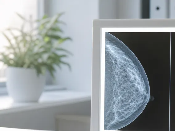
The LCIS medical condition is unique because it typically does not present with any noticeable symptoms. Unlike many breast conditions that may cause a lump, pain, or nipple discharge, LCIS is asymptomatic. This means it cannot be felt during a self-exam or a clinical breast exam, and it usually does not show up as a distinct mass on a mammogram.
Consequently, the LCIS definition and symptoms highlight that its discovery is almost always incidental. It is usually found when a biopsy is performed for another reason, such as an abnormal finding on a mammogram (e.g., microcalcifications or a benign-appearing mass) or a suspicious area identified during a physical examination. The diagnosis is confirmed through the microscopic examination of breast tissue by a pathologist.
Diagnosing and Treating LCIS
The diagnosis of LCIS primarily relies on a biopsy, which involves removing a small tissue sample from the breast for microscopic examination. This can be a core needle biopsy, performed with local anesthesia, or an excisional biopsy, which involves surgically removing a larger area of tissue. Pathological analysis of the tissue sample is crucial for confirming the presence of LCIS and differentiating it from other breast conditions.
Understanding LCIS diagnosis and treatment is important, as the management approach differs significantly from that of invasive breast cancer. Since LCIS itself is not an invasive cancer, it does not require treatments like chemotherapy or radiation therapy. Instead, the focus is on managing the elevated risk of developing future invasive breast cancer. Treatment strategies are tailored to the individual, considering factors such as personal risk profile, family history, and patient preferences.
Common management strategies for LCIS include:
- Enhanced Surveillance: This involves regular clinical breast exams, annual mammograms, and often supplemental imaging like breast MRI, particularly for women with additional risk factors.
- Risk-Reducing Medications: Medications such as tamoxifen or raloxifene may be prescribed to reduce the risk of developing invasive breast cancer. These drugs work by blocking estrogen’s effects on breast tissue.
- Surgical Excision: While not always necessary, surgical removal of the area containing LCIS may be recommended in specific cases, especially for certain subtypes like pleomorphic LCIS, or if the biopsy results are unclear.
LCIS Risk Factors and Prognosis
The primary significance of an LCIS diagnosis lies in its role as a strong indicator of increased future risk for developing invasive breast cancer. Women diagnosed with LCIS have an estimated 7-12 times higher risk of developing invasive breast cancer compared to women without LCIS, according to data from the American Cancer Society. This elevated risk applies to both breasts, meaning invasive cancer could develop in the breast where LCIS was found or in the opposite breast.
When considering LCIS risk factors and prognosis, it’s important to distinguish between factors that increase the risk of developing LCIS itself and factors that influence the progression from LCIS to invasive cancer. While the specific risk factors for developing LCIS are not fully understood, factors that may increase the likelihood of progressing to invasive breast cancer include a strong family history of breast cancer, the presence of certain genetic mutations (e.g., BRCA1/2), and the specific pathological features of the LCIS (e.g., pleomorphic LCIS). Age at diagnosis can also play a role, with younger women sometimes facing a higher relative risk.
The prognosis for LCIS itself is excellent because it is a non-invasive condition. However, the long-term outlook is closely tied to the increased risk of developing invasive breast cancer. Regular follow-up, adherence to screening guidelines, and participation in risk reduction strategies are crucial for women with LCIS. These measures help in early detection of any subsequent invasive cancer, significantly improving the chances of successful treatment and overall survival.



















