Diagnosis, Screening, and Early Detection of Pancreatic Cancer
Pancreatic cancer diagnosis usually involves a combination of imaging methods, laboratory testing, and clinical assessment to confirm the presence of a tumor and evaluate disease progression. Most cases involve pancreatic ductal adenocarcinoma (PDAC), although diagnostic approaches may differ for pancreatic neuroendocrine tumors and other rare pancreatic malignancies. Because symptoms often appear in advanced stages, early identification remains challenging. Accurate and timely diagnosis plays an important role in guiding treatment decisions and improving patient management.

Key Takeaways
- Early-stage pancreatic cancer often develops without clear symptoms.
- Imaging and biopsy are the most reliable diagnostic tools.
- Blood-based markers support evaluation but are not definitive.
- Screening is mainly recommended for individuals at elevated risk.
- Genetic testing is becoming increasingly important for identifying high-risk individuals and guiding treatment decisions.
Understanding Pancreatic Cancer Screening
Pancreatic cancer screening is not widely used for the general population due to the lack of highly sensitive and specific early detection tools. Instead, it is typically considered for individuals with a strong hereditary background or known genetic predispositions.
In high-risk groups, physicians may use imaging techniques such as MRI or endoscopic ultrasound for regular monitoring. These approaches can help identify abnormalities before symptoms develop. However, there is currently no standardized screening method that consistently detects early-stage disease in average-risk individuals.
How Doctors Approach Diagnosis
When symptoms or clinical findings suggest pancreatic cancer, doctors follow a structured evaluation process to determine how to diagnose pancreatic cancer accurately. This process usually includes reviewing the patient’s medical history, performing a physical examination, and selecting appropriate diagnostic tests.
Imaging Techniques
Imaging is central to diagnosing pancreatic cancer and helps determine the presence and extent of disease. Commonly used methods include:
- CT scans, especially pancreas-protocol CT, to visualize tumors and assess possible spread
- MRI scans for detailed soft tissue assessment
- Endoscopic ultrasound for high-resolution imaging of the pancreas and obtaining tissue samples when needed
These techniques provide essential information about tumor size, location, and involvement of surrounding tissues.
Biopsy Confirmation
A biopsy is often required to establish a definitive diagnosis. During this procedure, a small sample of tissue is collected and analyzed under a microscope to identify cancerous cells. This step is crucial for distinguishing malignant tumors from non-cancerous pancreatic conditions.
In clinical practice, biopsies are usually performed using image-guided approaches such as endoscopic ultrasound or CT guidance. These techniques improve accuracy and reduce procedural risks. The findings not only confirm the diagnosis but also provide insight into tumor type and cellular characteristics, which are important for further medical planning.
Laboratory Tests and Tumor Markers
Doctors frequently order pancreatic cancer test panels to evaluate overall health and detect abnormalities. These tests may include liver function tests and blood chemistry panels.
Blood-Based Indicators
A pancreatic cancer blood test can measure substances released by tumors into the bloodstream. One commonly used marker is CA 19-9.
The role of blood tests for pancreatic cancer is supportive rather than definitive. Elevated levels may indicate cancer, but they can also occur in non-cancerous conditions.
The marker cea pancreatic cancer is sometimes evaluated alongside CA 19-9 to provide additional context. However, it lacks specificity and cannot confirm the disease alone.
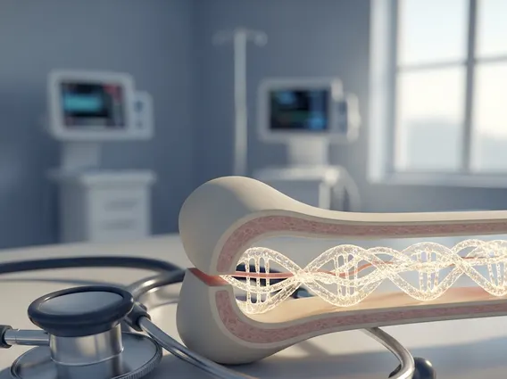
Genetic Testing and Risk Assessment
Advances in pancreatic cancer genetic testing are improving the ability to identify individuals at higher risk. These tests analyze inherited mutations that may increase susceptibility to pancreatic cancer.
Genetic testing is particularly useful for patients with a strong family history. It also helps guide personalized screening strategies and informs treatment decisions in some cases.
Early Detection Challenges
Efforts focused on pancreatic cancer early detection methods aim to identify the disease before it spreads. However, early-stage tumors often do not produce noticeable symptoms.
The concept of pancreatic cancer early detection remains an active area of research. Scientists are exploring biomarkers, imaging improvements, and artificial intelligence tools to enhance detection accuracy.
Common Diagnostic Tests Overview
The following table summarizes commonly used diagnostic tools:
| Test Type | Purpose | Limitations |
|---|---|---|
| CT Scan | Detects tumors and spread | May miss very small tumors |
| MRI | Provides detailed imaging | Higher cost and limited availability |
| Endoscopic Ultrasound | Close-up imaging and biopsy guidance | Invasive procedure |
| Blood Tests | Measures tumor markers and organ function | Not diagnostic alone |
| Biopsy | Confirms cancer diagnosis | Requires tissue sampling |
According to the National Cancer Institute, imaging combined with biopsy remains the gold standard for confirming pancreatic cancer diagnosis.
Role of ICD Coding and Clinical Documentation
Medical classification systems such as pancreatic cancer icd 10 codes are used to record diagnoses in a structured and internationally recognized format. These codes support administrative processes such as billing, but they also allow healthcare providers to organize patient data more effectively..
In addition, records related to family history of pancreatic cancer did 10 help clinicians identify patients who may carry a higher inherited risk. This information can influence follow-up strategies and determine whether closer monitoring is appropriate.
Visual Identification and Imaging Interpretation
Medical imaging often provides pictures of pancreatic cancer that help clinicians evaluate tumor characteristics. These images are analyzed by radiologists to detect abnormalities in the pancreas.
While imaging is essential, interpretation requires expertise to differentiate cancer from other pancreatic conditions such as inflammation or cysts.
Clinical Evaluation and Lab Work
Routine labs for pancreatic cancer may include liver enzyme tests, bilirubin levels, and complete blood counts. These evaluations help physicians understand how well the liver and other organs are functioning, especially since pancreatic tumors can block bile ducts and affect nearby structures. Abnormal results, such as elevated bilirubin, may indicate bile flow obstruction, which is sometimes associated with pancreatic tumors.
Although laboratory findings alone cannot confirm pancreatic cancer, they provide valuable clinical clues that support further investigation. When combined with imaging and physical examination, these results help doctors decide whether more advanced diagnostic procedures, such as specialized scans or biopsy, are necessary. This step-by-step approach ensures a more accurate and comprehensive evaluation.
Screening Strategies for High-Risk Individuals
Doctors determine how to test for pancreatic cancer in high-risk individuals based on genetic factors and family history. Screening programs often include periodic imaging and clinical evaluations.
Specialized centers may offer surveillance programs designed to detect early changes in the pancreas. These programs aim to improve outcomes through earlier intervention.
FAQs about Diagnosis, Screening, and Early Detection of Pancreatic Cancer
How is pancreatic cancer diagnosed?
Pancreatic cancer is diagnosed using a combination of imaging tests, laboratory evaluations, and biopsy confirmation. Doctors typically start with scans such as CT or MRI to detect abnormalities. If a suspicious mass is found, a biopsy is performed to confirm the presence of cancer cells. Blood tests may support the diagnosis but are not sufficient on their own.
Can ultrasound detect pancreatic cancer?
Ultrasound can sometimes detect pancreatic abnormalities, but it is not the most reliable method for diagnosing pancreatic cancer. Standard abdominal ultrasound may miss small tumors due to limited visibility. Endoscopic ultrasound is more accurate because it provides a closer and more detailed view of the pancreas and can also guide biopsy procedures.
Why is pancreatic cancer so hard to detect?
Pancreatic cancer is difficult to detect early because it often does not cause symptoms in its initial stages. The pancreas is located deep in the abdomen, making tumors harder to identify through routine exams. Additionally, early symptoms are vague and can resemble other common conditions, leading to delayed diagnosis.
Sources :