Inferior Vena Cava
The Inferior Vena Cava (IVC) is a vital blood vessel that plays a crucial role in the human circulatory system. Understanding its structure and function is essential for comprehending how deoxygenated blood returns to the heart from the lower parts of the body.
Key Takeaways
- The Inferior Vena Cava is the largest vein in the body, responsible for returning deoxygenated blood from the lower body to the right atrium of the heart.
- Its unique anatomical pathway involves traversing the abdomen and thorax, passing through the diaphragm.
- Proper inferior vena cava function is critical for maintaining cardiovascular health and preventing blood pooling in the lower extremities.
- Inferior vena cava syndrome occurs when blood flow through the IVC is obstructed, leading to a range of symptoms.
- Conditions affecting the IVC can have significant clinical implications, necessitating accurate diagnosis and management.
What is the Inferior Vena Cava (IVC)?
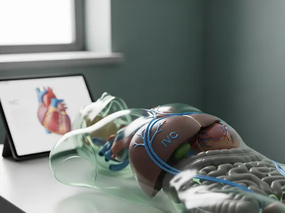
The Inferior Vena Cava (IVC) is the largest vein in the human body, serving as the primary conduit for deoxygenated blood returning to the heart from the lower half of the body. This crucial vessel collects blood from the legs, feet, abdomen, and pelvis, directing it upwards towards the right atrium. Its formation begins in the abdomen, where the two common iliac veins merge, and it ascends through the abdominal cavity and thorax before emptying into the heart. The IVC’s robust structure and strategic positioning are fundamental to its role in maintaining systemic circulation.
The Inferior Vena Cava is not merely a passive tube but an active participant in cardiovascular dynamics, influenced by respiratory movements and abdominal pressure changes. Its integrity is vital for preventing venous congestion and ensuring efficient blood return, which directly impacts cardiac output and overall circulatory efficiency.
Inferior Vena Cava Anatomy and Function
The inferior vena cava anatomy is complex and highly specialized to accommodate its function. It originates at the level of the fifth lumbar vertebra, formed by the union of the right and left common iliac veins. From there, it ascends along the right side of the vertebral column, posterior to the abdominal aorta. As it continues its upward course, it receives numerous tributaries, including the lumbar veins, renal veins (from the kidneys), hepatic veins (from the liver), and suprarenal veins (from the adrenal glands). This extensive network of tributaries ensures that deoxygenated blood from a vast region of the body is collected efficiently. The IVC then passes through the central tendon of the diaphragm at the level of the eighth thoracic vertebra before entering the right atrium of the heart.
The primary inferior vena cava function is to transport deoxygenated blood from the lower extremities and abdominopelvic organs back to the heart. This continuous return of blood is essential for the heart to pump oxygenated blood to the rest of the body. The IVC’s large diameter allows for a high volume of blood flow, and its relatively thin walls, compared to arteries, are characteristic of veins. While the IVC itself does not have valves to prevent backflow, the pressure gradient created by cardiac suction and respiratory movements helps propel blood towards the heart.
| Key Tributaries of the IVC | Drainage Area |
|---|---|
| Common Iliac Veins | Lower limbs and pelvis |
| Lumbar Veins | Posterior abdominal wall |
| Renal Veins | Kidneys |
| Hepatic Veins | Liver |
| Right Suprarenal Vein | Right adrenal gland |
| Right Gonadal Vein | Right testis/ovary |
Inferior Vena Cava Syndrome Explained
Inferior vena cava syndrome refers to a collection of signs and symptoms that arise when the blood flow through the IVC is obstructed or compressed. This obstruction can occur at any point along its course, from its formation in the pelvis to its entry into the heart. The causes of IVC syndrome are varied and can include external compression, such as from tumors (e.g., liver cancer, renal cell carcinoma), enlarged lymph nodes, or pregnancy. Internal obstruction can result from blood clots (thrombosis), often associated with conditions like deep vein thrombosis (DVT) extending into the IVC, or from congenital abnormalities like IVC webs or atresia.
The symptoms of inferior vena cava syndrome depend on the location and severity of the obstruction. Common manifestations include swelling (edema) in the legs, ankles, and feet, which may be unilateral or bilateral. Patients may also experience distended superficial veins in the abdomen and lower back, as blood tries to find collateral pathways. Other symptoms can include pain or discomfort in the abdomen or back, kidney dysfunction if the obstruction affects renal vein drainage, and liver congestion with elevated liver enzymes if hepatic veins are involved. In severe cases, reduced cardiac output and hypotension can occur due to impaired venous return.
Diagnosis typically involves imaging studies such as ultrasound, CT scans, or MRI, which can visualize the IVC and identify the cause of obstruction. Treatment strategies vary widely based on the underlying cause and may include anticoagulation for blood clots, surgical removal of tumors, angioplasty and stenting to open narrowed segments, or placement of an IVC filter to prevent pulmonary embolism in specific high-risk situations. Early diagnosis and intervention are crucial to prevent complications and improve patient outcomes.