Stage 0 Transitional Cell Carcinoma In Situ Of The Renal Pelvis And Ureter
Stage 0 Transitional Cell Carcinoma In Situ Of The Renal Pelvis And Ureter represents a very early and non-invasive form of cancer affecting the lining of the kidney’s urine-collecting system and the tube connecting it to the bladder.
Key Takeaways
- Stage 0 Transitional Cell Carcinoma In Situ is a non-invasive, high-grade cancer confined to the surface lining of the renal pelvis or ureter.
- Symptoms are often subtle, with painless blood in the urine (hematuria) being the most common indicator.
- Diagnosis relies on a combination of imaging, endoscopic procedures, and tissue biopsy.
- Treatment aims to remove cancerous cells while preserving the affected organ, often through minimally invasive techniques.
- The prognosis is generally excellent due to its early stage, but long-term surveillance is crucial due to the risk of recurrence.
What is Stage 0 Transitional Cell Carcinoma In Situ Of The Renal Pelvis And Ureter?
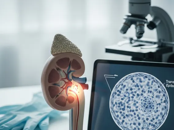
Stage 0 Transitional Cell Carcinoma In Situ Of The Renal Pelvis And Ureter refers to a non-invasive, high-grade cancer that originates in the urothelium, the specialized lining of the urinary tract. Specifically, this condition affects the renal pelvis, which is the funnel-shaped part of the kidney that collects urine, and/or the ureter, the tube that carries urine from the kidney to the bladder. The term “in situ” signifies that the abnormal cancer cells are confined strictly to the innermost layer of the lining and have not invaded deeper into the muscle or connective tissues of the organ. This early stage means the cancer has not spread, making it highly treatable.
This form of cancer is considered high-grade, meaning the cells appear significantly abnormal under a microscope and have a higher potential for progression if left untreated. However, its non-invasive nature at Stage 0 is a critical factor in its favorable prognosis. Understanding its precise location and non-invasive status is key to effective management and surveillance.
Recognizing Symptoms and Diagnosing Stage 0 Transitional Cell Carcinoma In Situ
Recognizing transitional cell carcinoma in situ symptoms can be challenging, as the condition often presents with subtle or no noticeable signs in its earliest stages. The most common symptom is painless hematuria, which is the presence of blood in the urine. This blood may be visible to the naked eye (gross hematuria) or only detectable under a microscope (microscopic hematuria). Other less common and non-specific symptoms might include mild flank pain or changes in urinary frequency, but these are rarely the primary indicators for Stage 0 disease.
Due to the often subtle nature of symptoms, diagnosis typically involves a combination of advanced imaging and endoscopic procedures. These methods allow healthcare providers to visualize the urinary tract directly and obtain tissue samples for definitive analysis. Key diagnostic steps include:
- Urinalysis and Urinary Cytology: To detect blood in the urine and examine urine cells for any abnormalities or cancerous changes.
- Imaging Studies: Such as CT Urogram or MRI, which provide detailed images of the kidneys, renal pelvis, and ureters to identify any suspicious lesions or blockages.
- Cystoscopy and Ureteroscopy: These are endoscopic procedures where a thin, flexible tube with a camera is inserted through the urethra to visualize the bladder (cystoscopy) and, if necessary, advanced into the ureters and renal pelvis (ureteroscopy) to directly inspect the lining.
- Biopsy: During ureteroscopy, suspicious areas can be biopsied, providing tissue samples that are then examined by a pathologist to confirm the diagnosis of transitional cell carcinoma in situ and determine its grade.
Treatment Approaches and Prognosis for Stage 0 TCC of the Renal Pelvis and Ureter
The primary goal of stage 0 TCC renal pelvis treatment options is to eradicate the cancerous cells while preserving the kidney and ureter function whenever possible. Given the non-invasive nature of Stage 0 disease, treatment often focuses on minimally invasive techniques. The specific approach depends on factors such as the size and location of the lesion, as well as the patient’s overall health.
Common treatment strategies include:
- Endoscopic Resection or Ablation: Using a ureteroscope, the visible tumor can be precisely removed or destroyed using laser ablation or electrocautery. This approach is often preferred for smaller, localized lesions.
- Intracavitary Therapy: This involves instilling chemotherapy agents (e.g., mitomycin C) or immunotherapy agents (e.g., Bacillus Calmette-Guérin or BCG) directly into the renal pelvis or ureter. This topical treatment aims to kill remaining cancer cells and reduce the risk of recurrence, particularly for multifocal or diffuse in situ lesions.
- Surveillance: For very small, low-risk lesions or after initial treatment, a rigorous surveillance program involving regular imaging and endoscopic examinations is crucial to monitor for recurrence.
- Nephroureterectomy: In rare cases, if conservative treatments are unsuccessful, the disease is extensive, or there is a high risk of progression, surgical removal of the affected kidney and entire ureter may be considered. However, this is less common for true Stage 0 in situ disease.
The prognosis stage 0 transitional cell carcinoma is generally excellent due to its early detection and non-invasive nature. With appropriate treatment, cure rates are very high. However, patients with transitional cell carcinoma in situ have a significant risk of recurrence, not only at the original site but also elsewhere in the urinary tract, including the bladder. Therefore, long-term, diligent surveillance is essential to detect any new or recurrent lesions promptly and ensure the best possible long-term outcomes.