Solar Keratosis
Solar Keratosis is a common skin condition resulting from prolonged sun exposure, primarily affecting areas frequently exposed to ultraviolet (UV) radiation. It is considered a precancerous lesion, highlighting the importance of early detection and treatment to prevent potential progression.
Key Takeaways
- Solar Keratosis is a rough, scaly patch of skin caused by chronic sun damage.
- It is considered a precancerous lesion, meaning it has the potential to develop into squamous cell carcinoma.
- Common symptoms include dry, rough, or scaly patches, often appearing on sun-exposed areas like the face, scalp, hands, and arms.
- Treatment options range from topical medications to cryotherapy, photodynamic therapy, and surgical removal.
- Regular skin examinations and consistent sun protection are crucial for prevention and early management of the condition.
What is Solar Keratosis?
Solar Keratosis refers to a common skin condition characterized by rough, scaly patches that develop on the skin after years of exposure to the sun’s ultraviolet (UV) rays. These lesions are typically small, ranging from a few millimeters to a centimeter or more, and can vary in color from flesh-toned to pink, red, or brown. They often feel like sandpaper to the touch and are most commonly found on sun-exposed areas such as the face, ears, lips, bald scalp, neck, back of hands, and forearms.
While often benign, solar keratoses are considered precancerous lesions, meaning they have the potential to evolve into a type of skin cancer called squamous cell carcinoma. Early identification and treatment are therefore essential to manage the risk of progression. The condition is more prevalent in older adults, individuals with fair skin, and those with a history of significant sun exposure or sunburns.
Solar Keratosis: Symptoms, Causes, and Cancer Risk
Understanding the manifestations and origins of this condition is crucial for effective management. The primary solar keratosis symptoms and causes are directly linked to sun exposure and how the skin reacts to UV radiation over time. Symptoms often appear gradually and can include:
- Rough, dry, or scaly patches of skin, often feeling like sandpaper.
- Patches that are flesh-colored, pink, red, or brownish.
- Itching, burning, or tenderness in the affected area.
- A hard, wart-like surface in some cases.
- Lesions that are typically less than 1 inch (2.5 cm) in diameter.
The main cause of solar keratosis is chronic exposure to ultraviolet (UV) radiation from sunlight or tanning beds. UV radiation damages the keratinocytes, which are the main cells in the outermost layer of the skin (epidermis). Over time, this damage leads to abnormal cell growth and the characteristic lesions. Risk factors include fair skin, blue or light-colored eyes, red or blond hair, a history of frequent sun exposure or sunburns, and a weakened immune system.
Regarding the question, is solar keratosis skin cancer? Solar Keratosis itself is not skin cancer, but rather a precancerous condition. It is classified as an early form of squamous cell carcinoma (SCC) confined to the outermost layer of the skin. While most solar keratoses do not progress to invasive cancer, a small percentage can. According to the American Academy of Dermatology (AAD), approximately 5-10% of solar keratoses may eventually develop into squamous cell carcinoma, which is the second most common type of skin cancer. Therefore, monitoring and treating these lesions are important steps in preventing skin cancer.
Solar Keratosis Treatment Options
A variety of solar keratosis treatment options are available, chosen based on the number, location, and severity of the lesions, as well as the patient’s overall health and preferences. The goal of treatment is to remove or destroy the affected skin cells, thereby reducing the risk of progression to squamous cell carcinoma. Common treatments include:
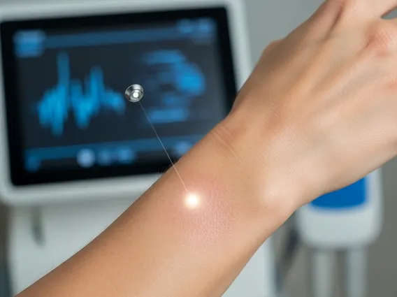
One of the most frequently used methods is cryotherapy, where liquid nitrogen is applied to freeze and destroy the affected skin cells. This procedure is quick and effective for individual lesions. Topical medications, such as 5-fluorouracil (5-FU) cream, imiquimod cream, or diclofenac gel, can be prescribed for widespread or multiple lesions. These medications work by causing the abnormal cells to die and peel off over several weeks.
Other treatment modalities include photodynamic therapy (PDT), which involves applying a light-sensitizing solution to the skin, followed by exposure to a special light that activates the solution to destroy the lesions. Chemical peels can also be used to remove the top layers of skin, including the solar keratoses. In some cases, surgical removal (shave excision or curettage and electrodesiccation) may be necessary for larger or more suspicious lesions. Regular follow-up with a dermatologist is crucial after treatment to monitor for new lesions or recurrence.