Scanner
A Scanner in a medical context refers to a sophisticated diagnostic imaging device used to visualize internal structures of the body. These devices play a crucial role in modern medicine, aiding in the detection, diagnosis, and monitoring of various diseases and conditions.
Key Takeaways
- Medical scanners are essential diagnostic tools that create detailed images of the body’s internal structures.
- They are utilized for a wide range of applications, including disease detection, injury assessment, and guiding medical procedures.
- Scanners operate using diverse physical principles, such as X-rays, magnetic fields, or sound waves, to generate images.
- Common types include CT, MRI, Ultrasound, and PET, each offering unique insights into different tissues and physiological processes.
- The choice of scanner depends on the specific diagnostic need, as each technology provides distinct advantages for various medical conditions.
What is a Scanner: Purpose and Applications
A medical Scanner is an advanced piece of equipment designed to produce images of the inside of the human body without invasive surgery. This capability is fundamental to modern diagnostics, allowing healthcare professionals to observe organs, tissues, and skeletal structures in detail. The primary purpose of these devices is to assist in the accurate diagnosis of medical conditions, monitor disease progression, and evaluate the effectiveness of treatments.
Scanners are indispensable tools across numerous medical specialties. They are used for a wide array of applications, from detecting subtle abnormalities to providing real-time guidance during complex procedures. For instance, they can identify tumors, assess bone fractures, visualize blood flow, and examine soft tissues like the brain or heart. This broad utility makes them cornerstones in fields such as oncology, cardiology, neurology, and orthopedics.
How Does a Scanner Work?
The fundamental principle behind how a medical scanner works involves the interaction of various forms of energy with the body’s tissues, followed by the detection and processing of the resulting signals into an image. Each type of scanner employs a distinct energy source and detection method. For example, Computed Tomography (CT) scanners use X-rays, while Magnetic Resonance Imaging (MRI) scanners utilize powerful magnetic fields and radio waves. Ultrasound devices, on the other hand, rely on high-frequency sound waves.
Regardless of the specific technology, the general process involves three key stages: energy emission, interaction with the body, and signal detection. The scanner emits energy that passes through or reflects off the body. Different tissues absorb, reflect, or alter this energy in unique ways. Detectors then capture these altered signals, which are subsequently sent to a computer. Sophisticated algorithms process these signals to reconstruct detailed cross-sectional or three-dimensional images of the internal anatomy, allowing clinicians to visualize structures and identify anomalies.
Types of Scanners and Their Technology
There are several distinct types of medical scanners, each employing different physical principles to generate images and providing unique diagnostic insights. Understanding these various types of scanners explained helps in appreciating their specific applications in clinical practice. The overarching scanner technology overview reveals a diverse landscape of imaging modalities.
Here are some of the most common types of medical scanners:
- Computed Tomography (CT) Scanner: Utilizes X-rays rotated around the patient to create cross-sectional images. These images are then combined by a computer to produce detailed slices of bones, blood vessels, and soft tissues. CT scans are particularly effective for quickly examining people who may have internal injuries from car accidents or other types of trauma.
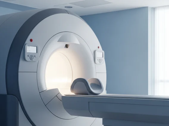
- Magnetic Resonance Imaging (MRI) Scanner: Employs strong magnetic fields and radio waves to generate detailed images of organs and soft tissues. Unlike CT scans, MRI does not use ionizing radiation, making it suitable for repeated imaging. It excels in visualizing the brain, spinal cord, joints, and internal organs.
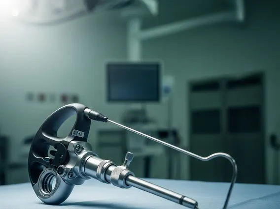
- Ultrasound Scanner: Uses high-frequency sound waves to create real-time images of internal body structures. A transducer emits sound waves that bounce off tissues and organs, and the echoes are then converted into an image. Ultrasound is commonly used for fetal imaging, abdominal examinations, and evaluating blood flow.
- Positron Emission Tomography (PET) Scanner: Detects gamma rays emitted by a small amount of radioactive tracer injected into the patient. The tracer accumulates in areas of high metabolic activity, such as cancerous cells, allowing PET scans to reveal the functional activity of organs and tissues rather than just their structure.
Each scanner type offers distinct advantages depending on the clinical question. For instance, CT is often preferred for bone injuries and acute trauma, while MRI provides superior detail for soft tissue pathologies. Ultrasound is excellent for dynamic imaging and is radiation-free, making it safe for pregnancy. PET scans are invaluable for oncology, neurology, and cardiology to assess metabolic function. The continuous advancement in scanner technology continues to enhance diagnostic capabilities, leading to more precise and timely medical interventions.