
Gerotas Fascia
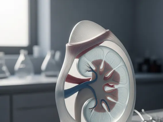
Gerota’s fascia is a fibrous connective tissue envelope surrounding the kidney and adrenal gland, playing a critical role in both normal renal anatomy and the clinical staging of kidney cancer.
Key Takeaways
- Gerota’s fascia is a multilayered fascial sheath enclosing the kidney, adrenal gland, and perinephric fat.
- It serves as a natural anatomical barrier that helps contain infection, inflammation, and tumor spread.
- Invasion beyond Gerota’s fascia by renal cell carcinoma (RCC) is a defining criterion for T4 disease in TNM staging.
- T4 classification indicates locally advanced disease and significantly influences treatment decisions and prognosis.
- Surgeons rely on fascial boundaries during nephrectomy to achieve complete tumor resection.
Gerota’s Fascia: Anatomy and Function
Gerota’s fascia is a bilaminar fibrofatty sheath located in the retroperitoneal space. It consists of an anterior layer (sometimes called the prerenal fascia) and a posterior layer (retrorenal fascia), both of which fuse superiorly around the adrenal gland. Laterally, the two layers merge to form the lateroconal fascia, while inferiorly they remain open or loosely fused, allowing potential communication with the pelvic extraperitoneal space. The structure encloses the kidney, adrenal gland, and surrounding perinephric fat within a defined compartment.
The primary functions of this fascial layer include:
- Containment: Limits the spread of perinephric infections, hematomas, and inflammatory processes.
- Structural support: Maintains the positional stability of the kidney within the retroperitoneum.
- Surgical landmark: Guides surgeons during radical and partial nephrectomy by defining resection planes.
The Gerotas fascia anatomy and clinical significance extend beyond simple containment. Because the fascial sheath forms a relatively resistant boundary, it temporarily restricts the outward migration of pathological processes — a property that becomes especially relevant in oncologic settings. The thickness and integrity of the fascia can vary between individuals, but its spatial relationships with adjacent structures remain consistent enough to serve as a reliable anatomical reference in both imaging and surgery.
Clinical Significance of Gerota’s Fascia in Renal Cell Carcinoma Staging
Renal cell carcinoma (RCC) is the most common malignant tumor of the kidney in adults, accounting for approximately 90% of kidney cancers. According to the American Cancer Society, an estimated 81,000 new cases of kidney cancer are diagnosed annually in the United States, with RCC comprising the vast majority. The anatomical boundaries defined by the fascia are central to how these tumors are staged and treated.
The Gerotas fascia role in kidney cancer staging is codified in the TNM (Tumor, Node, Metastasis) classification system published by the American Joint Committee on Cancer (AJCC). Under this system, tumor invasion beyond the fascia into adjacent organs or the ipsilateral adrenal gland by direct extension defines pT4 disease — the most locally advanced primary tumor category. This distinction carries meaningful prognostic weight, as T4 tumors are associated with substantially lower five-year survival rates compared to organ-confined disease.
| TNM Stage | Relationship to Gerota’s Fascia | Clinical Implication |
|---|---|---|
| T1–T2 | Tumor confined within the fascia | Localized disease; surgery often curative |
| T3 | Tumor extends into perinephric fat but not beyond the fascia | Locally advanced; radical nephrectomy indicated |
| T4 | Tumor invades beyond the fascia | Highest local stage; multimodal therapy considered |
In the context of Gerotas fascia in renal cell carcinoma, preoperative cross-sectional imaging — particularly contrast-enhanced CT and MRI — is used to assess whether the fascial margin appears intact or breached. Radiologists evaluate fascial thickening, stranding of perinephric fat, and direct organ invasion as indicators of extrafascial spread. These imaging findings directly inform surgical planning and help determine eligibility for nephron-sparing approaches versus radical resection.
Intraoperatively, surgeons use the fascia as the outer boundary of the resection specimen during radical nephrectomy, aiming to remove the kidney, surrounding fat, and adrenal gland en bloc within the intact fascial envelope. Maintaining this boundary intact reduces the risk of positive surgical margins and local recurrence. When the fascia is already breached by tumor, the surgical challenge increases considerably, often requiring resection of adjacent structures and consideration of systemic therapy.



















