Radical Local Excision
Radical Local Excision is a surgical procedure designed to remove a cancerous or precancerous lesion along with a surrounding margin of healthy tissue. This approach aims to ensure the complete removal of abnormal cells, thereby minimizing the risk of recurrence.
Key Takeaways
- Radical Local Excision is a surgical technique for removing tumors with clear margins.
- It is primarily indicated for early-stage cancers and certain high-risk lesions.
- The procedure involves excising the lesion and a surrounding area of healthy tissue.
- Pathological examination of the removed tissue is crucial to confirm clear margins.
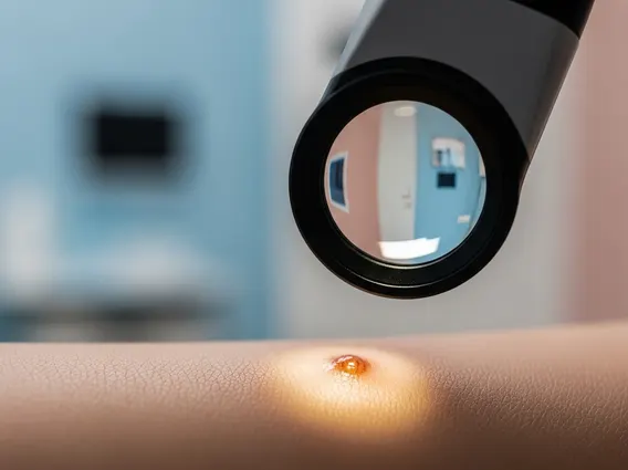
What is Radical Local Excision?
Radical Local Excision refers to a surgical operation where a tumor or lesion is removed with a significant margin of surrounding healthy tissue. The term “radical” in this context emphasizes the intent to achieve complete removal by taking a wider margin than a simple excision, aiming to eliminate any microscopic extensions of the disease that might not be visible to the naked eye. The primary goal is to achieve “clear margins,” meaning that no cancer cells are found at the edges of the removed tissue when examined under a microscope.
The radical local excision meaning is rooted in oncology principles, where the thorough removal of primary tumors is paramount to preventing local recurrence and improving patient outcomes. This meticulous approach is particularly vital for certain types of cancers where local control is a key determinant of prognosis. By removing a sufficient amount of healthy tissue around the lesion, surgeons aim to maximize the chances of eradicating all cancerous cells.
Indications for Radical Local Excision
Radical Local Excision is typically indicated for specific types of cancers and high-risk lesions where complete removal with clear margins is critical. The decision to perform this procedure depends on several factors, including the type, size, location, and stage of the tumor, as well as the patient’s overall health. Common indications include:
- Early-stage melanoma and other aggressive skin cancers.
- Certain soft tissue sarcomas.
- Some types of breast cancer, particularly for smaller tumors or as part of breast-conserving surgery.
- High-grade dysplastic lesions or carcinoma in situ in various anatomical sites.
The procedure is often chosen when there is a high risk of local recurrence if the lesion is not completely removed, or when the tumor has aggressive features that necessitate a wider margin. Pathological assessment prior to surgery, often through a biopsy, helps determine the appropriate surgical approach and the extent of the margin required.
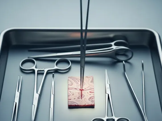
The Radical Local Excision Procedure
The radical local excision procedure involves several distinct stages, beginning with careful pre-operative planning. This includes imaging studies and biopsies to precisely map the lesion and assess its depth and spread. On the day of the surgery, the patient receives appropriate anesthesia, which can range from local anesthesia with sedation to general anesthesia, depending on the size and location of the lesion and patient preference.
The surgeon then meticulously excises the lesion, ensuring that a predetermined margin of healthy tissue is removed around and beneath it. This excised tissue is immediately sent to a pathology lab for examination. In some cases, a “frozen section” analysis may be performed during the surgery to rapidly check the margins for clear cells. If cancer cells are found at the margins, the surgeon may remove additional tissue until clear margins are achieved. Once the excision is complete and clear margins are confirmed, the wound is closed using sutures, staples, or sometimes a skin graft or flap, especially for larger defects.
This radical local excision surgery is a cornerstone in the treatment of many localized cancers. Post-operative care typically involves pain management, wound care, and monitoring for any signs of infection or complications. Patients are usually followed up to ensure proper healing and to monitor for any signs of recurrence, often involving regular clinical examinations and imaging studies. The success of the procedure relies heavily on achieving clear margins, which significantly reduces the likelihood of the cancer returning at the surgical site.