Intraoperative Ultrasound
Intraoperative Ultrasound (IOU) is an advanced medical imaging technique that provides real-time visualization of anatomical structures and pathologies during surgical procedures. This technology significantly enhances a surgeon’s ability to make precise decisions and optimize patient outcomes.
Key Takeaways
- Intraoperative Ultrasound (IOU) is a real-time imaging modality used directly in the operating room to guide surgical interventions.
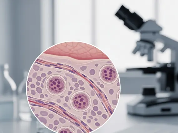
- It employs high-frequency sound waves to create detailed images, allowing surgeons to visualize tissues and abnormalities not visible to the naked eye.
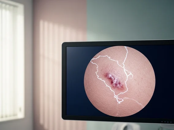
- IOU enhances surgical precision by providing immediate feedback on tumor margins, lesion localization, and critical anatomical structures.
- The technology offers significant intraoperative ultrasound benefits, including improved patient safety, reduced complication rates, and more complete resections.
- Its intraoperative ultrasound uses span various specialties, from neurosurgery and liver resections to vascular and gynecological procedures.
What is Intraoperative Ultrasound (IOU)?
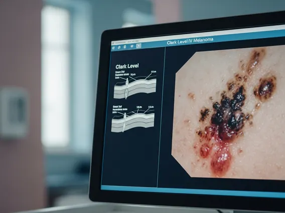
Intraoperative Ultrasound (IOU) refers to the use of ultrasound imaging directly within the operating room environment to assist surgeons during complex procedures. Unlike preoperative imaging, IOU provides dynamic, real-time views of internal structures, allowing for immediate assessment and guidance. This capability is crucial for identifying lesions, delineating tumor margins, and navigating around vital anatomical features that might be obscured by blood or tissue during an open or minimally invasive surgery. The immediate feedback loop provided by IOU helps surgeons confirm the extent of disease, verify the completeness of resections, and avoid damage to surrounding healthy tissue.
The integration of ultrasound technology into surgical practice has revolutionized precision in many fields. It allows for a more tailored and accurate approach to surgery, moving beyond what can be seen with the naked eye or felt by hand. This real-time imaging is particularly valuable in scenarios where the target tissue is soft, mobile, or difficult to distinguish from normal anatomy, providing an invaluable tool for surgical decision-making.
How Intraoperative Ultrasound Works
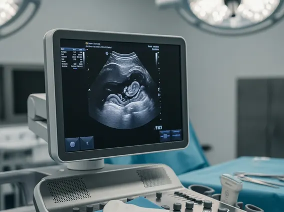
The fundamental principle behind how intraoperative ultrasound works involves the emission and reception of high-frequency sound waves. A specialized transducer, often sterilely draped, is placed directly on the organ or tissue being operated on. This transducer generates sound waves that travel into the body, reflect off different tissue densities, and then return to the transducer. The time it takes for these echoes to return, along with their intensity, is processed by a computer to create a real-time, two-dimensional image displayed on a monitor.
The key to IOU’s effectiveness lies in its ability to provide immediate visual updates. As the surgeon operates, they can continuously scan the area, adjusting their approach based on the live images. This dynamic interaction allows for precise localization of tumors, identification of vascular structures, and confirmation of complete removal of diseased tissue. Modern IOU systems often include advanced features like Doppler imaging to assess blood flow, contrast-enhanced ultrasound to highlight specific lesions, and 3D reconstruction capabilities, further enhancing their diagnostic and guidance utility.
Clinical Applications and Benefits of IOU
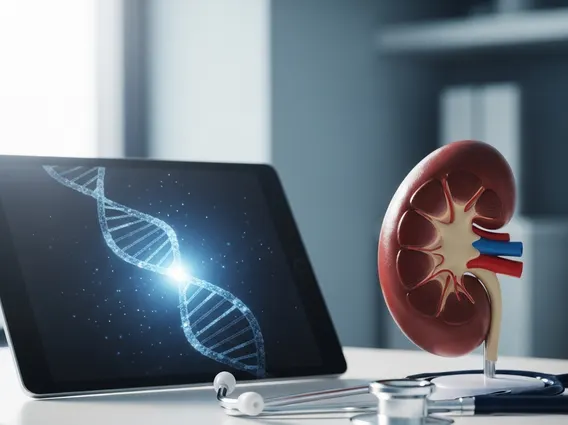
The intraoperative ultrasound uses are extensive and continue to expand across various surgical disciplines due to its versatility and real-time capabilities. In neurosurgery, IOU is invaluable for guiding tumor resections, especially in eloquent brain areas, by precisely delineating tumor boundaries and confirming the extent of removal. For liver surgery, it aids in localizing small or deep lesions, mapping vascular anatomy, and ensuring complete tumor ablation or resection, which is critical for preventing recurrence. Other significant applications include vascular surgery for identifying plaques and guiding stent placement, urology for kidney and prostate procedures, and gynecology for ovarian and uterine surgeries.
The intraoperative ultrasound benefits for both surgeons and patients are substantial. These include:
- Enhanced Precision: Provides real-time, high-resolution images, allowing surgeons to operate with greater accuracy and confidence.
- Improved Tumor Resection: Helps achieve more complete removal of cancerous or diseased tissue, potentially reducing the need for repeat surgeries.
- Reduced Complications: Facilitates the identification and avoidance of critical structures like nerves and blood vessels, thereby minimizing surgical risks.
- Real-time Feedback: Offers immediate visual confirmation of surgical progress, enabling on-the-spot adjustments to the operative plan.
- Preservation of Healthy Tissue: Allows for more targeted interventions, sparing healthy surrounding tissue and potentially leading to faster recovery times.
According to a review published in the journal Ultrasound in Medicine & Biology, the use of IOU has been associated with improved surgical outcomes and reduced recurrence rates in several oncological procedures. This highlights its critical role in modern surgical practice, contributing to safer and more effective patient care.