Sentinel Lymph Node Mapping
Sentinel Lymph Node Mapping is a vital diagnostic procedure in oncology, used to assess the spread of cancer and guide subsequent treatment decisions. This technique helps identify the specific lymph nodes most likely to contain cancer cells that have metastasized from a primary tumor.

Key Takeaways
- Sentinel Lymph Node Mapping identifies the first lymph nodes to which cancer cells are likely to spread from a primary tumor.
- The procedure is crucial for accurate cancer staging and helps determine the most appropriate treatment plan.
- It involves injecting a tracer (radioactive substance or blue dye) near the tumor to locate the sentinel nodes.
- By precisely identifying affected nodes, it often allows for less extensive surgery, reducing the risk of complications like lymphedema.
- This technique is a standard of care for several cancers, including melanoma and breast cancer.
What is Sentinel Lymph Node Mapping and Why is it Performed?
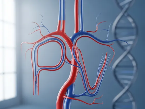
Sentinel Lymph Node Mapping is a surgical technique designed to identify the sentinel lymph node(s), which are the initial lymph nodes where cancer cells are most likely to spread from a primary tumor. These nodes act as a “first line of defense” in the lymphatic system, filtering fluid and potentially trapping cancer cells before they can disseminate further.
The primary purpose of sentinel lymph node mapping is to accurately determine if cancer has spread beyond its original site into the lymphatic system. This information is critical for precise cancer staging, which, in turn, dictates the most effective treatment strategy. By identifying only the potentially affected nodes, this procedure can often prevent the need for a more extensive lymph node dissection, a surgery that removes many lymph nodes and carries a higher risk of side effects such as lymphedema (swelling caused by fluid buildup).
According to the National Cancer Institute, sentinel lymph node biopsy is a standard of care for many early-stage cancers, including breast cancer and melanoma, which collectively affect hundreds of thousands of individuals annually in the U.S. alone. Its widespread adoption underscores its importance in minimizing surgical invasiveness while providing essential prognostic information.
The Sentinel Lymph Node Mapping Procedure Explained
The sentinel lymph node mapping procedure is a precise surgical technique typically performed before or during the surgery to remove the primary tumor. The process involves several key steps to accurately locate the sentinel lymph node(s).
The procedure generally follows these steps:
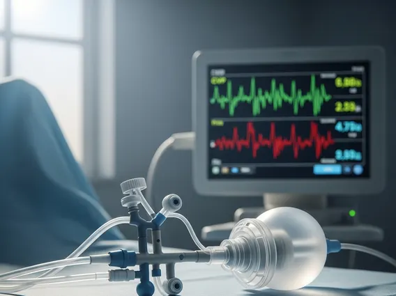
- Tracer Injection: A small amount of a radioactive tracer (radioisotope) and/or a blue dye is injected into the tissue surrounding the primary tumor.
- Lymphatic Flow: The injected tracer then travels through the lymphatic vessels, following the same path that cancer cells would take, to the first lymph nodes in the drainage basin—the sentinel nodes.
- Detection: During surgery, the surgeon uses a specialized handheld gamma probe to detect the radioactivity and/or visually identifies the blue-stained lymph nodes. These are the sentinel nodes.
- Surgical Removal: The identified sentinel lymph node(s) are carefully removed through a small incision.
- Pathological Examination: The removed nodes are immediately or subsequently sent to a pathology lab, where they are meticulously examined under a microscope for the presence of cancer cells.
This process, often referred to as a sentinel lymph node biopsy explained, offers a less invasive alternative to a complete lymph node dissection. If the sentinel nodes are found to be free of cancer cells, it strongly suggests that the cancer has not spread to other lymph nodes, potentially sparing the patient from further, more extensive surgery and its associated complications. Conversely, if cancer cells are detected in the sentinel nodes, it indicates that the cancer has begun to spread, necessitating further treatment which may include additional lymph node removal, radiation therapy, or systemic treatments.