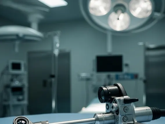
Thoracoscope
A thoracoscope is a vital medical instrument used in minimally invasive procedures to examine and treat conditions within the chest cavity. This advanced device allows medical professionals to gain direct visualization of the pleura, lungs, and other thoracic structures without the need for large incisions.
Key Takeaways
- A thoracoscope is a slender, illuminated instrument used for minimally invasive examination and treatment of the chest cavity.
- It consists of a camera, light source, working channels, and an eyepiece, enabling detailed visualization and surgical intervention.
- Thoracoscopy, the procedure using a thoracoscope, is crucial for diagnosing and treating various lung and pleural conditions.
- Common uses include biopsy, fluid drainage, pleurodesis, and staging of certain cancers.
- This technique offers benefits such as reduced pain, shorter hospital stays, and quicker recovery compared to traditional open surgery.
What is a Thoracoscope?
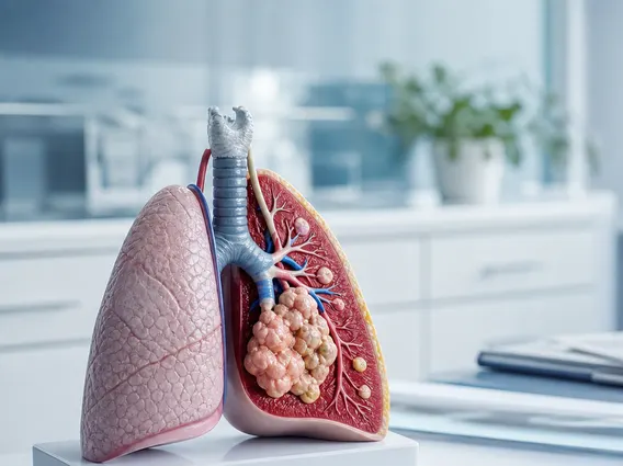
A thoracoscope is a specialized endoscope designed to be inserted into the chest cavity, or thorax, through small incisions. This instrument provides direct visual access to the pleural space, lungs, mediastinum, and other thoracic structures. Its primary function is to facilitate diagnostic examinations and therapeutic interventions in a minimally invasive manner. The use of a thoracoscope allows clinicians to accurately assess conditions, collect tissue samples, and perform various procedures with enhanced precision and reduced patient discomfort compared to traditional open surgical methods.
Components and Function of a Thoracoscope
A thoracoscope operates by combining several key components to provide visualization and enable intervention within the chest. The device typically consists of a rigid or flexible tube equipped with a high-resolution camera or optical fibers, a powerful light source, and one or more working channels. The camera transmits real-time images to a monitor, allowing the medical team to observe the internal structures in detail. The light source illuminates the surgical field, ensuring clear visibility. The working channels accommodate various surgical instruments, such as forceps for biopsies, scissors for tissue dissection, or suction devices for fluid removal. This integrated design allows for precise manipulation and intervention within the confined space of the chest cavity.
Thoracoscope Uses and Associated Procedures
Thoracoscopy explained refers to the medical procedure where a thoracoscope is used to visualize the chest cavity and perform diagnostic or therapeutic interventions. This minimally invasive approach is widely utilized for a range of pulmonary and pleural conditions. The procedure offers significant advantages over traditional open chest surgery, including smaller incisions, reduced pain, shorter hospital stays, and faster recovery times for patients.
Common procedures performed using a thoracoscope include:
- Pleural Biopsy: Obtaining tissue samples from the pleura (the lining of the lungs and chest wall) to diagnose conditions like mesothelioma, tuberculosis, or other inflammatory diseases.
- Fluid Drainage: Removing excess fluid from the pleural space (pleural effusion) that can cause breathing difficulties, often for diagnostic analysis or symptom relief.
- Pleurodesis: A procedure to prevent recurrent pleural effusions by introducing a substance that causes the lung to adhere to the chest wall.
- Lung Biopsy: Taking tissue samples from the lung itself to diagnose interstitial lung diseases, infections, or suspected malignancies.
- Staging of Lung Cancer: Assessing the extent of lung cancer spread to lymph nodes or other structures within the chest, which is crucial for treatment planning.
- Treatment of Pneumothorax: Repairing air leaks and preventing recurrent collapsed lung episodes.
These applications highlight the versatility and importance of thoracoscopy in modern respiratory medicine, providing a less invasive alternative for managing complex thoracic conditions.