Thermography
Thermography is a non-invasive diagnostic tool that captures and analyzes heat patterns emitted from the body’s surface. This advanced imaging technique provides valuable insights into physiological processes, making it a significant asset in various medical and clinical fields.
Key Takeaways
- Thermography is a non-invasive imaging technique measuring surface temperature variations.
- It works by detecting infrared radiation emitted by the body, converting it into a visual thermal map.
- Applications include assessing inflammation, nerve function, vascular conditions, and musculoskeletal injuries.
- Benefits include its non-contact nature, lack of radiation exposure, and detection of subtle physiological changes.
- It serves as a complementary diagnostic tool, providing functional information.
What is Thermography?
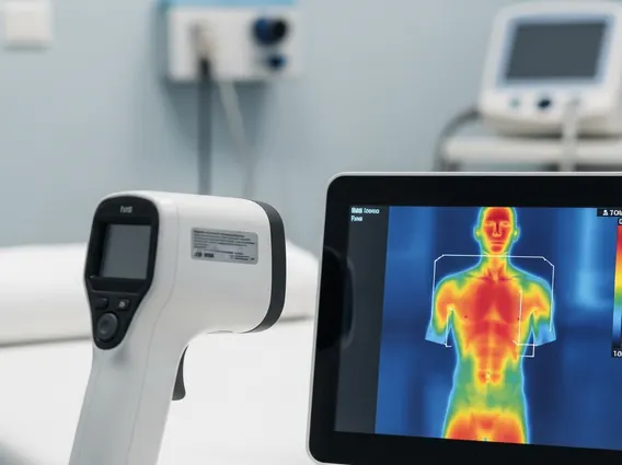
Thermography refers to a non-invasive imaging technique that measures and records the temperature of the skin’s surface. This method utilizes specialized infrared cameras to detect the heat emitted by the body, a byproduct of metabolic activity and blood flow. The resulting thermal images, or thermograms, display temperature variations as different colors or shades, allowing clinicians to visualize areas of abnormal heat distribution. For instance, increased heat can indicate inflammation or heightened metabolic activity, while decreased heat might suggest reduced blood flow or nerve dysfunction. This capability makes it a valuable tool for understanding physiological processes without direct contact or radiation exposure.
How Thermography Works
The principle behind how does thermography work? involves the detection of infrared radiation. Every object with a temperature above absolute zero emits infrared energy, including the human body. Thermographic cameras are equipped with highly sensitive infrared detectors that capture this emitted energy. These detectors convert the infrared radiation into electrical signals, which are then processed by a computer to create a real-time visual representation of temperature distribution across the body’s surface. The software assigns different colors to different temperatures, typically with warmer colors (like red and orange) representing higher temperatures and cooler colors (like blue and purple) indicating lower temperatures. This process allows for precise mapping of thermal patterns, revealing subtle temperature asymmetries indicative of underlying physiological changes.
The accuracy of thermographic imaging relies on factors such as camera resolution, environmental control, and patient preparation. Standardized protocols ensure consistent and reliable results, enhancing the interpretation of thermograms for diagnostic purposes. The technology provides a functional assessment, complementing structural imaging techniques like X-rays or MRI by offering insights into physiological activity.
Applications and Benefits of Thermography
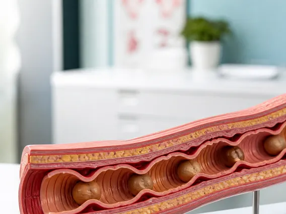
Thermography applications explained cover a broad spectrum within the medical field, primarily focusing on conditions characterized by changes in surface temperature. One significant area is the assessment of musculoskeletal injuries, where inflammation or nerve impingement can lead to detectable thermal patterns. It is also used in the evaluation of chronic pain syndromes, such as fibromyalgia and complex regional pain syndrome (CRPS), by identifying areas of abnormal sympathetic nervous system activity. Furthermore, thermography can assist in diagnosing vascular disorders, including peripheral vascular disease, by visualizing areas of compromised blood flow.
Other notable applications include:
- Neurological Assessments: Identifying nerve root irritation or peripheral neuropathy.
- Rheumatological Conditions: Monitoring inflammatory processes in conditions like arthritis.
- Breast Health: As an adjunctive tool to mammography, detecting thermal asymmetries.
- Sports Medicine: Pinpointing areas of strain or injury for targeted treatment.
The benefits of thermography are numerous, making it an attractive option for both patients and clinicians. Firstly, it is completely non-invasive and non-contact, ideal for sensitive areas or patients with pain. Secondly, it involves no radiation exposure, making it safe for repeated use, even in pregnant women or children. Thirdly, thermography provides a physiological assessment, offering insights into functional changes that might precede anatomical alterations visible on other imaging modalities. This early detection capability can be crucial for timely intervention. While thermography is a valuable diagnostic adjunct, it provides supportive information and does not replace conventional medical treatments or established diagnostic procedures. Always consult with a qualified healthcare professional for diagnosis and treatment recommendations.