Diagnosis, Screening, and Early Detection of Pheochromocytoma
Pheochromocytoma is a rare neuroendocrine tumor, primarily of the adrenal glands, that can cause severe health issues due to excessive hormone release. Timely pheochromocytoma diagnosis is critical for preventing life-threatening complications and ensuring effective treatment.
Key Takeaways
- Pheochromocytomas are rare tumors producing excess catecholamines, leading to symptoms like high blood pressure, headaches, and palpitations.
- Early detection is vital to prevent serious cardiovascular and neurological complications.
- Screening guidelines for pheochromocytoma target individuals with specific risk factors or suggestive symptoms.
- Best tests for pheochromocytoma diagnosis include biochemical tests (urine and blood metanephrines) followed by imaging studies.
- Prompt medical consultation upon recognizing early signs of pheochromocytoma is crucial for effective management.
Understanding Pheochromocytoma and Its Diagnosis
Gaining a comprehensive understanding of pheochromocytoma is the first step toward effective management and treatment. This rare condition, though challenging to diagnose, is treatable when identified early.
What is Pheochromocytoma?
A pheochromocytoma is a rare tumor that originates in the chromaffin cells of the adrenal glands, which are located on top of the kidneys. These tumors secrete excessive amounts of catecholamines—hormones like adrenaline (epinephrine) and noradrenaline (norepinephrine)—leading to a range of symptoms. While most pheochromocytomas are benign, they can cause significant health problems if left undiagnosed. Understanding what is pheochromocytoma and how is it diagnosed is essential for patients and healthcare providers alike.
Why Early Diagnosis Matters
The chronic or episodic release of high levels of catecholamines can lead to severe and potentially life-threatening complications, including hypertensive crises, stroke, heart attack, and kidney damage. Without early intervention, these conditions can be fatal. For instance, untreated pheochromocytoma can lead to a mortality rate of up to 50% in some cases due to cardiovascular events, according to studies published in the Journal of Clinical Endocrinology & Metabolism. Therefore, a prompt and accurate pheochromocytoma diagnosis is paramount to mitigate these risks, stabilize the patient’s condition, and allow for timely surgical removal, which is often curative.
Recognizing Early Signs of Pheochromocytoma
The symptoms of pheochromocytoma can be varied and often mimic other conditions, making early recognition challenging. However, certain patterns of symptoms should raise suspicion.
Common Symptoms to Watch For
Recognizing the early signs of pheochromocytoma is crucial for timely medical evaluation. Symptoms often arise from the surge of catecholamines and can be paroxysmal (occurring in attacks) or sustained. The classic triad of symptoms includes episodic headaches, sweating, and heart palpitations, often accompanied by severe high blood pressure (hypertension). Other common manifestations may include anxiety, tremors, pallor, and abdominal pain. These symptoms can be non-specific, making diagnosis challenging, but their recurrent or severe nature warrants investigation, especially if they are unexplained or resistant to standard treatments.
When to Seek Medical Advice
Individuals experiencing recurrent episodes of severe headaches, unexplained sweating, rapid heartbeats, or sudden spikes in blood pressure should seek medical advice promptly. This is especially true if these symptoms occur together, are increasing in frequency or severity, or are resistant to conventional hypertension treatments. While these symptoms can be indicative of many conditions, discussing them with a healthcare professional is essential to determine if further investigation for conditions like pheochromocytoma is warranted.
Pheochromocytoma Screening Guidelines
Given the rarity of pheochromocytoma, screening is typically recommended for specific high-risk populations rather than the general public.
Who Needs Screening?
Screening guidelines for pheochromocytoma are primarily focused on identifying individuals at higher risk, as the condition is relatively rare in the general population, with an estimated incidence of 0.8 per 100,000 person-years, according to the National Cancer Institute. High-risk groups include those with a family history of pheochromocytoma or related genetic syndromes such as Multiple Endocrine Neoplasia type 2 (MEN2), Von Hippel-Lindau (VHL) disease, or Neurofibromatosis type 1 (NF1). Patients with resistant hypertension, unexplained hypertensive crises, or an adrenal incidentaloma (a mass found on the adrenal gland during imaging for another reason) also warrant consideration for screening. Understanding pheochromocytoma screening ensures that at-risk individuals receive appropriate evaluation.
Screening Procedures Explained
Initial screening for pheochromocytoma typically involves biochemical tests designed to detect elevated levels of catecholamines or their metabolites. These tests are non-invasive and serve as the first line of investigation for suspected cases. They help to identify the biochemical evidence of the tumor’s activity, guiding subsequent diagnostic steps and helping clinicians determine the likelihood of a pheochromocytoma before proceeding to more complex imaging studies.
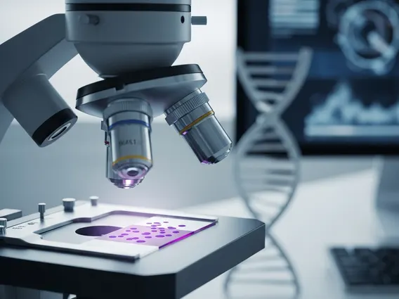
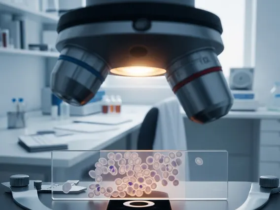
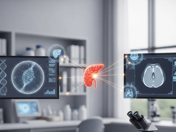
Key Diagnostic Tests for Pheochromocytoma
Accurate diagnosis of pheochromocytoma relies on a combination of biochemical confirmation and precise tumor localization through imaging.
Biochemical Testing: Urine and Blood
The pheochromocytoma diagnosis methods rely heavily on biochemical confirmation of excess catecholamine production. The best tests for pheochromocytoma diagnosis involve measuring metanephrines and normetanephrines, which are breakdown products of adrenaline and noradrenaline, respectively. These can be measured in a 24-hour urine collection or in plasma (blood). Plasma free metanephrines are often considered highly sensitive for screening, while 24-hour urinary fractionated metanephrines and catecholamines are also very effective. These tests are crucial for confirming the biochemical presence of the tumor, with specific cut-off values indicating a high probability of pheochromocytoma. Proper patient preparation, including avoiding certain medications and foods, is vital to prevent false positive results.
Imaging Studies: Locating the Tumor
Once biochemical tests confirm the presence of excess catecholamines, imaging studies are used to pinpoint the tumor’s location. This is a critical step in how to detect pheochromocytoma early and plan for surgical removal. Common imaging modalities include:
- Computed Tomography (CT) scans: Provide detailed anatomical images of the adrenal glands and abdomen, effectively identifying most adrenal tumors.
- Magnetic Resonance Imaging (MRI): Offers excellent soft tissue contrast, particularly useful for adrenal masses and in cases where radiation exposure is a concern, such as in children or pregnant women.
- Metaiodobenzylguanidine (MIBG) scintigraphy: A nuclear medicine scan that uses a radioactive tracer taken up by chromaffin cells, helping to identify pheochromocytomas, especially those outside the adrenal glands (paragangliomas) or metastatic disease.
- Positron Emission Tomography (PET) scans: With specific tracers, PET scans can also be highly effective in locating tumors, particularly in complex or recurrent cases, offering functional information about the tumor’s metabolic activity.
The choice of imaging depends on individual patient factors, the suspected location of the tumor, and the specific clinical scenario.
Frequently Asked Questions
What are the most common symptoms of pheochromocytoma?
The most common symptoms of pheochromocytoma include episodic headaches, excessive sweating, and heart palpitations, often accompanied by severe and sudden increases in blood pressure. Other signs can involve anxiety, tremors, pallor, and abdominal pain. These symptoms occur due to the tumor’s release of high levels of stress hormones, known as catecholamines. If you experience these symptoms regularly or severely, especially together, it is important to consult a healthcare professional for evaluation.
How is pheochromocytoma typically diagnosed?
Pheochromocytoma diagnosis typically begins with biochemical tests to measure catecholamines and their metabolites (metanephrines) in 24-hour urine collections or plasma. Elevated levels suggest the presence of the tumor. Following biochemical confirmation, imaging studies such as CT scans, MRI, or MIBG scintigraphy are performed to locate the tumor, which is most commonly found in the adrenal glands. These steps are crucial for accurate identification and treatment planning.
Who should be screened for pheochromocytoma?
Screening guidelines for pheochromocytoma recommend testing for individuals with a family history of pheochromocytoma or related genetic syndromes (e.g., MEN2, VHL, NF1). Additionally, patients with incidentally discovered adrenal masses, resistant hypertension, or unexplained hypertensive crises should be screened. Early screening in these high-risk groups is vital for preventing serious complications associated with delayed diagnosis and ensuring timely intervention.