
Urachus
The urachus is a fascinating remnant of fetal development, playing a crucial role during gestation but typically becoming non-functional after birth. Understanding its anatomy and potential anomalies is vital in medical contexts.
Key Takeaways
- The urachus is a fibrous cord connecting the fetal bladder to the umbilical cord, essential for waste removal during development.
- Normally, it obliterates after birth, transforming into the median umbilical ligament.
- Failure of the urachus to close can lead to various anomalies, including urachal cysts, patent urachus, urachal sinuses, and diverticula.
- Symptoms of urachal anomalies often involve abdominal pain, umbilical discharge, or urinary issues.
- Treatment typically involves surgical intervention to remove the abnormal urachal tissue and prevent complications.
What is the Urachus: Anatomy and Function
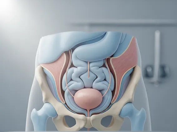
Urachus refers to a fibrous remnant of the allantois, a structure present during embryonic development. In the fetus, it is a tubular connection between the apex of the bladder and the umbilical cord. Its primary function during gestation is to channel urine from the fetal bladder to the allantois, which then empties into the amniotic fluid. This process is crucial for fetal waste excretion before the kidneys are fully mature and functional. Urachal anomalies are relatively rare, occurring in approximately 1 in 5,000 to 1 in 10,000 live births, though many remain asymptomatic and undiagnosed.
After birth, the urachus normally undergoes obliteration, meaning it closes off and transforms into a solid cord. This cord is known as the median umbilical ligament, which extends from the apex of the bladder to the umbilicus. The complete closure of the urachus is a vital developmental step, as its failure to do so can lead to various congenital anomalies. Understanding urachus anatomy and function is essential for diagnosing and treating these conditions, which can manifest in different forms depending on the extent and location of the persistent opening.
Urachal Cyst and Patent Urachus: Symptoms and Treatment
When the urachus fails to completely obliterate after birth, it can result in several anomalies, each with distinct clinical presentations. One common anomaly is an urachal cyst, which forms when both ends of the urachus close, but the middle portion remains patent and fills with fluid. These cysts are often asymptomatic but can become infected, leading to a range of symptoms.
Common urachal cyst symptoms include:
- Abdominal pain, typically localized in the lower abdomen
- A palpable mass or swelling near the umbilicus
- Fever and localized tenderness if the cyst becomes infected
- Umbilical discharge if the cyst ruptures or forms a sinus to the skin
- Urinary symptoms like painful urination or frequent urges if the cyst irritates the bladder
Another significant anomaly is a patent urachus, where the entire urachus remains open, creating a direct connection between the bladder and the umbilicus. This condition allows urine to continuously drain from the umbilicus, leading to persistent wetness and irritation around the navel. The causes of a patent urachus are congenital, stemming from the failure of the urachal lumen to close during fetal development. This persistent connection poses a risk for infection and skin irritation.
The patent urachus causes treatment approach primarily involves surgical correction. For a patent urachus, surgical excision of the entire tract, including a small cuff of bladder, is typically performed to prevent recurrent infections and urine leakage. Similarly, symptomatic urachal cysts, especially those that are infected or large, also require surgical removal. This usually involves excising the cyst and any associated urachal remnant. Early diagnosis and intervention are crucial to prevent complications such as recurrent infections, stone formation within the urachal remnant, or, rarely, malignant transformation, although the latter is extremely rare.


