
Upper Gi Series
The Upper Gi Series is a diagnostic imaging procedure used to examine the upper part of the gastrointestinal tract. This article will explain what the procedure entails, why it is performed, and what patients can expect during preparation and the examination itself.
Key Takeaways
- An Upper Gi Series uses X-rays and a contrast material to visualize the esophagus, stomach, and duodenum.
- It helps diagnose conditions like ulcers, reflux, strictures, and tumors.
- Preparation involves fasting and avoiding certain medications.
- The procedure is generally safe and provides valuable diagnostic information.
What is an Upper GI Series?
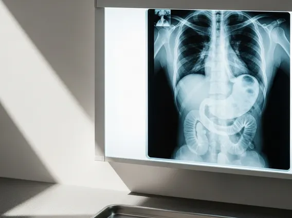
What is an Upper Gi Series refers to a specialized X-ray examination that allows doctors to visualize the upper part of the gastrointestinal (GI) tract. This includes the esophagus, stomach, and the first part of the small intestine, known as the duodenum. During the procedure, the patient drinks a liquid contrast material, typically barium, which coats the lining of these organs, making them visible on X-ray images. This allows radiologists to observe the shape, function, and any abnormalities within these structures.
The procedure is also sometimes referred to as a barium swallow or an esophagram, particularly when the focus is primarily on the esophagus. It provides dynamic images, meaning the radiologist can watch the barium move through the digestive tract in real-time, helping to identify issues with swallowing or the movement of food.
Why an Upper GI Series is Performed
An Upper Gi Series is performed to help diagnose a range of conditions affecting the esophagus, stomach, and duodenum. Doctors recommend this procedure when patients experience symptoms that suggest problems in these areas. Understanding why get an upper gi series is crucial for patients experiencing persistent digestive issues.
Common reasons for undergoing this examination include:
- Difficulty swallowing (dysphagia)
- Persistent heartburn or acid reflux (gastroesophageal reflux disease, GERD)
- Unexplained nausea, vomiting, or abdominal pain
- Blood in stool or unexplained weight loss, which could indicate ulcers, inflammation, or tumors
- Suspected hernias, strictures (narrowing), or blockages in the upper GI tract
According to the American College of Radiology, an Upper Gi Series can be particularly useful in identifying structural abnormalities that might not be easily seen with other diagnostic methods, providing a clear visual assessment of the upper digestive system. For instance, it can detect peptic ulcers, which affect approximately 10% of people in Western countries at some point in their lives (Source: National Institute of Diabetes and Digestive and Kidney Diseases – NIDDK).
Upper GI Series Procedure and Preparation
The upper gi series procedure explained involves several steps to ensure clear imaging and patient comfort. Before the procedure begins, patients will receive specific instructions for upper gi series preparation.
Preparation
Proper preparation is vital for accurate results. Typically, patients are instructed to:
- Fast for several hours (usually 8-12 hours) before the examination, meaning no food or drink.
- Avoid smoking or chewing gum during the fasting period, as these can stimulate digestive juices and interfere with the barium coating.
- Inform their doctor about any medications they are taking, as some may need to be temporarily stopped.
- Discuss any known allergies, especially to contrast materials.
Procedure
During the procedure, the patient will typically stand or lie on an X-ray table. The radiologist or technologist will ask them to drink the barium solution. The barium has a chalky taste but is generally tolerable. As the patient swallows, the radiologist will take a series of X-ray images or a video (fluoroscopy) to observe the barium’s movement through the esophagus, stomach, and duodenum. The patient may be asked to change positions or hold their breath briefly to help capture different views. Sometimes, a gas-producing agent is given to distend the stomach, providing even clearer images. The entire procedure usually takes about 30-60 minutes. After the examination, patients are encouraged to drink plenty of fluids to help flush the barium from their system and prevent constipation.


